
Cerebral aqueduct: Difference between revisions
Content deleted Content added
| Line 48: | Line 48: | ||
==Additional images== |
==Additional images== |
||
<gallery> |
<gallery> |
||
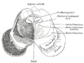
Image:Gray710.png|Transverse section through [[mid-brain]]. |
Image:Gray710.png|Transverse section through [[mid-brain]]; number 2 indicates the cerebral aqueduct. |
||
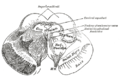
Image:Gray711.png|Transverse section of mid-brain at level of inferior colliculi. |
Image:Gray711.png|Transverse section of mid-brain at level of inferior colliculi. |
||
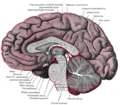
Image:Gray712.png|Transverse section of mid-brain at level of superior colliculi. |
Image:Gray712.png|Transverse section of mid-brain at level of superior colliculi. |
||
Revision as of 20:02, 31 August 2011
| Cerebral aqueduct | |
|---|---|
 Section through superior colliculus showing path of oculomotor nerve. | |
 Drawing of a cast of the ventricular cavities, viewed from the side. | |
| Details | |
| Identifiers | |
| Latin | aqueductus mesencephali (cerebri) |
| MeSH | D002535 |
| NeuroNames | 509 |
| NeuroLex ID | birnlex_1261 |
| TA98 | A14.1.06.501 |
| TA2 | 5910 |
| FMA | 78467 |
| Anatomical terms of neuroanatomy | |
The mesencephalic duct, also known as the aqueductus mesencephali, aqueduct of Sylvius or the cerebral aqueduct, contains cerebrospinal fluid (CSF), is within the mesencephalon (or midbrain) and connects the third ventricle in the diencephalon to the fourth ventricle, which is between the pons and cerebellum.
Development
The cerebral aqueduct, similarly to other parts of the ventricular system of the brain, develops from the central canal of the neural tube. Specifically, the duct originates from the portion of the neural tube that is present in the developing mesencephalon, hence the name "mesencephalic duct." [1]
Pathology
A blockage in this duct is a cause of hydrocephalus.
See also
References
- ^ Le, Tao; Bhushan, Vikas; Vasan, Neil (2010). First Aid for the USMLE Step 1: 2010 20th Anniversary Edition. USA: The McGraw-Hill Companies, Inc. p. 126. ISBN 978-0-07-163340-6.
External links
- Atlas image: n2a3p2 at the University of Michigan Health System
Additional images
-
 Transverse section through mid-brain; number 2 indicates the cerebral aqueduct.
 -
 Transverse section of mid-brain at level of inferior colliculi.
 -
 Transverse section of mid-brain at level of superior colliculi.
 -
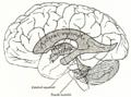
 Median sagittal section of brain.
 -
 Scheme showing relations of the ventricles to the surface of the brain.
 -
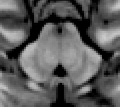 MRI section of mid-brain


This neuroanatomy article is a stub. You can help Wikipedia by expanding it. |