Cloacal membrane: Difference between revisions
No edit summary |
Rescuing 1 sources and tagging 0 as dead. #IABot (v1.5beta) |
||
| Line 29: | Line 29: | ||
* {{EmbryologyUNC|genital|021}} |
* {{EmbryologyUNC|genital|021}} |
||
* [http://embryology.med.unsw.edu.au/Medicine/images/stage11cloacal.jpg Diagram at unsw.edu.au] |
* [http://embryology.med.unsw.edu.au/Medicine/images/stage11cloacal.jpg Diagram at unsw.edu.au] |
||
* [http://www.ana.ed.ac.uk/anatomy/humat/notes/embryo/urogenital/cloacal.htm Overview at ana.ed.ac.uk] |
* [https://archive.is/20031119143454/http://www.ana.ed.ac.uk/anatomy/humat/notes/embryo/urogenital/cloacal.htm Overview at ana.ed.ac.uk] |
||
{{Development of urinary and reproductive systems}} |
{{Development of urinary and reproductive systems}} |
||
Revision as of 15:55, 9 August 2017
| Cloacal membrane | |
|---|---|
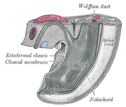
 Tail end of human embryo from fifteen to eighteen days old. | |
| Details | |
| Carnegie stage | 7 |
| Days | 15 |
| Precursor | caudal end of the primitive streak |
| Identifiers | |
| Latin | membrana cloacalis |
| TE | membrane_by_E5.4.0.0.0.0.15 E5.4.0.0.0.0.15 |
| Anatomical terminology | |
The cloacal membrane is the membrane that covers the embryonic cloaca during the development of the urinary and reproductive organs.
It is formed by ectoderm and endoderm coming into contact with each other.[1] As the human embryo grows and caudal folding continues, the urorectal septum divides the cloaca into a ventral urogenital sinus and dorsal anorectal canal. Before the urorectal septum has an opportunity to fuse with the cloacal membrane, the membrane ruptures, exposing the urogenital sinus and dorsal anorectal canal to the exterior. Later on, an ectodermal plug, the anal membrane, forms to create the lower third of the rectum.
References
![]() This article incorporates text in the public domain from page 47 of the 20th edition of Gray's Anatomy (1918)
This article incorporates text in the public domain from page 47 of the 20th edition of Gray's Anatomy (1918)
External links
- Swiss embryology (from UL, UB, and UF) hdisqueembry/triderm04
- genital-021—Embryo Images at University of North Carolina
- Diagram at unsw.edu.au
- Overview at ana.ed.ac.uk
