
Septum pellucidum
| Septum pellucidum | |
|---|---|
 Scheme of rhinencephalon. (Septum pellucidum visible at top center.) | |
 Median sagittal section of brain. (Septum pellucidum visible at center.) | |
| Identifiers | |
| MeSH | D012688 |
| NeuroNames | 256 |
| NeuroLex ID | nlx_144186 |
| TA98 | A14.1.09.262 |
| TA2 | 5647 |
| FMA | 61844 |
| Anatomical terms of neuroanatomy | |
The septum pellucidum (also called the septum lucidum), and not to be confused with the medial septum, is a thin, triangular, vertical membrane separating the anterior horns of the left and right lateral ventricles of the brain. It runs as a sheet from the corpus callosum down to the fornix.
Layers
The septum pellucidum actually consists of two layers or laminae of both white and gray matter,[1] called the laminae septi pellucidi. During fetal development there is a space between the two laminae called the cavum septum pellucidum which, in ninety per cent of cases, disappears during infancy.[2][3] The cavum is occasionally referred to as the fifth ventricle but the term has lost favor in recent years, as the space is usually not continuous with the ventricular system and does not contain cerebrospinal fluid.[4] Indeed fifth ventricle has been used for other purposes in recent years.[5]
Location
The septum pellucidum is located in the midline of the brain, between the two cerebral hemispheres. It is attached inferior to the corpus callosum, the large collection of nerve fibers that connect the two cerebral hemispheres. It is attached to the anterior part of the fornix, and on either side of the structure are the two lateral ventricles.
Confusion over the term
The septum pellucidum is often confused with the septal nuclei. Logically, the septum pellucidum is a septum in the medial plane and could therefore be termed 'medial septum', but this is incorrect. The term medial septum is reserved for a small group of nuclei which are closely associated with the septum pellucidum.
Pathology
Absence of the septum pellucidum or corpus callosum, caused by mutations in the HESX1 gene, is associated with septo-optic dysplasia. This may result in hypothalamic dysfunction and hypopituitarism, as well as problems of vision, coordination, and intelligence, as well as other unusual symptoms.
Additional images
-
 Mesal aspect of a brain sectioned in the median sagittal plane.
 -
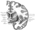
 Horizontal section of right cerebral hemisphere.
 -
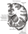 Coronal section through anterior cornua of lateral ventricles.
 -
 Coronal section of brain through anterior commissure.
 -
 The fornix and corpus callosum from below.
 -
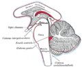 Diagram showing the positions of the three principal subarachnoid cisternæ.


References
- ^ Lennart Heimer; Gary W. Van Hoesen (16 November 2007). Anatomy of neuropsychiatry: the new anatomy of the basal forebrain and its implications for neuropsychiatric illness. Academic Press. p. 28. ISBN 9780123742391. Retrieved 24 December 2010.
- ^ David L. Clark; Nash N. Boutros; Mario F. Mendez (2010). The Brain and Behavior: An Introduction to Behavioral Neuroanatomy. Cambridge University Press. pp. 217–8. ISBN 9780521142298. Retrieved 24 December 2010.
- ^ Love J, Hollenhorst R (1956). "Bilateral palsy of the sixth cranial nerve caused by a cyst of the septum pellucidum (fifth ventricle) and cured by pneumoencephalography". Mayo Clin Proc. 31 (2): 43–6. PMID 13289891.
- ^ Alonso J, Coveñas R, Lara J, Piñuela C, Aijón J (1989). "The cavum septi pellucidi: a fifth ventricle?". Acta Anat (Basel). 134 (4): 286–90. doi:10.1159/000146704. PMID 2741657.
{{cite journal}}: CS1 maint: multiple names: authors list (link) - ^ Liccardo G, Ruggeri F, De Cerchio L, Floris R, Lunardi P (2005). "Fifth ventricle: an unusual cystic lesion of the conus medullaris". Spinal Cord. 43 (6): 381–4. doi:10.1038/sj.sc.3101712. PMID 15655569.
{{cite journal}}: CS1 maint: multiple names: authors list (link)