Stapes
| Stapes | |
|---|---|
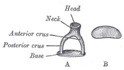
 A. Left stapes. B. Base of stapes, medial surface. | |
 | |
| Details | |
| Precursor | 2nd branchial arch[1] |
| Identifiers | |
| Latin | stapellos |
| MeSH | D013199 |
| TA98 | A15.3.02.033 |
| TA2 | 895 |
| FMA | 52751 |
| Anatomical terms of bone | |
The stapes is a bone in the middle ear of humans and other mammals which is involved in the conduction of sound vibrations to the inner ear. The the stirrup-shaped small bone is one of three ossicle in the middle ear. The stapes receives vibrations from the incus, to which it is connected laterally, and transmits these to the oval window, medially. The stapes is the smallest and lightest bone in the human body, and is so-called because of its resemblance to a stirrup (Latin: Stapes).
Structure
The stapes is the third bone of the three ossicles in the middle ear. The stapes is a stirrup-shaped bone, and the smallest in the human body. It lies directly on the oval window. The stapes is described as having a base, resting on the oval window, as well as a head that articulates with the incus. These are connected by anterior and posterior limbs (Latin: crura).[2]: 862 The stapes articulates with the incus through the incudostapedial joint. [3]
Development
The stapes develops from the second pharyngeal arch during the 6th to 8th week of embryological life. The central cavity of the stapedius is due to the presence embryologically of the stapedial artery, which later regresses. [4][3]
In animals
The stapes is one of three ossicles in mammals. In non-mammalian four-legged animalss, the bone homologous to the stapes is usually called the columella; however, in reptiles, either term may be used. In fish, the homologous bone is called the hyomandibular, and is part of the gill arch supporting either the spiracle or the jaw, depending on species. The equivalent term in amphibians is the pars media plectra. [3] [5]: 481–482
Function
Situated between the malleolus and the incus, the stapes transmits the sound vibrations to the incus to the membrane of the inner ear inside the oval window. The stapes is also stabilized by the stapedius muscle, which is innervated by the facial nerve.[6]
Clinical relevance
Otosclerosis is a congenital or chronic state causing ossification of the bone. This causes the stapes to become adherent to the oval window, and is a cause of conductive hearing loss. A stapedectomy or stapedotomy may be carried to rectify this.
The stapedial artery persists in about 1 in 15,000 persons.
History
The stapes is commonly described as having been discovered the professor Giovanni Filippo Ingrassia in 1546 at the University of Naples,[7] although this remains the nature of some controversy. [8] The bone is so-named because of its resemblance to a stirrup (Latin: Stapes), an example of a late Latin word, probably created in mediaeval times from "to stand" (Latin: stapia), as stirrups did not exist in the early Latin-speaking world. [9]
-
The stapes, as first described by Giovanni Filippo Ingrassia.
Additional images
-
Stapes and 10 cent euro coin
-
CT image of the stapes.
-
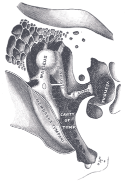
Auditory ossicles. Tympanic cavity. Deep dissection.
See also
References
- ^ hednk-023—Embryo Images at University of North Carolina
- ^ Drake, Richard L. (2005). Gray's anatomy for students. Philadelphia: Elsevier/Churchill Livingstone. ISBN 978-0-8089-2306-0.
{{cite book}}: Unknown parameter|coauthors=ignored (|author=suggested) (help) - ^ a b c Chapman, SC (2011 Jan 1). "Can you hear me now? Understanding vertebrate middle ear development". Frontiers in bioscience (Landmark edition). 16: 1675–92. PMID 21196256.
{{cite journal}}: Check date values in:|date=(help) - ^ Rodriguez-Vazquez, J. F. (2005). "Development of the stapes and associated structures in human embryos". Journal of Anatomy. 207 (2): 165–173. doi:10.1111/j.1469-7580.2005.00441.x.
{{cite journal}}: Unknown parameter|month=ignored (help) - ^ Romer, Alfred Sherwood; Parsons, Thomas S (1977). The Vertebrate Body. Philadelphia, PA: Holt-Saunders International. ISBN 0-03-910284-X.
- ^ "Dissector Answers - Ear & Nasal Cavity". University of Michigan. Retrieved January 2010.
{{cite web}}: Check date values in:|accessdate=(help) - ^ Dispenza, F (2013 Oct). "The discovery of stapes". Acta otorhinolaryngologica Italica : organo ufficiale della Societa italiana di otorinolaringologia e chirurgia cervico-facciale. 33 (5): 357–9. PMID 24227905.
{{cite journal}}: Check date values in:|date=(help); Unknown parameter|coauthors=ignored (|author=suggested) (help) - ^ Mudry, Albert (2013). "Disputes Surrounding the Discovery of the Stapes in the Mid 16th Century". Otology & Neurotology. 34 (3): 588–592. doi:10.1097/MAO.0b013e31827d8abc.
{{cite journal}}: Unknown parameter|month=ignored (help) - ^ Harper, Douglas. "Stapes (n.)". Online Etymology Dictionary. Retrieved 27 December 2013.
- Scanning electron microscopy images and energy-dispersive X-ray microanalysis of the stapes in otosclerosis and van der Hoeve syndrome. Vol. 110. 2000 Sep. pp. 1505–10.
{{cite book}}:|journal=ignored (help); Check date values in:|date=(help); Unknown parameter|authors=ignored (help)
External links
- "Development of mammal skull from primitive reptile skull". archive.org. Archived from the original on 2006-11-11. Retrieved January 2010.
{{cite web}}: Check date values in:|accessdate=(help) - "Reptilia Lab". Juniata Collage. Retrieved January 2010.
{{cite web}}: Check date values in:|accessdate=(help) - "3-D Virtual Models of the Human Temporal Bone and Related Structures". Eaton Peabody Laboratory of Auditory Physiology. Retrieved January 2010.
{{cite web}}: Check date values in:|accessdate=(help)


