Systematic evolution of ligands by exponential enrichment

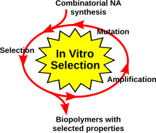
Systematic Evolution of Ligands by Exponential Enrichment (SELEX), also referred to as in vitro selection or in vitro evolution, is a combinatorial chemistry technique in molecular biology for producing oligonucleotides of either single-stranded DNA or RNA that specifically bind to a target ligand or ligands.[1][2][3] Although SELEX has emerged as the most commonly used name for the procedure, some researchers have referred to it as SAAB (selected and amplified binding site) and CASTing (Cyclic amplification and selection of targets)[4][5]
The process begins with the synthesis of a very large oligonucleotide library consisting of randomly generated sequences of fixed length flanked by constant 5' and 3' ends that serve as primers. For a randomly generated region of length n, the number of possible sequences in the library is 4n (n positions with four possibilities (A,T,C,G) at each position). The sequences in the library are exposed to the target ligand - which may be a protein or a small organic compound - and those that do not bind the target are removed, usually by affinity chromatography. The bound sequences are eluted and amplified by PCR to prepare for subsequent rounds of selection in which the stringency of the elution conditions is increased to identify the tightest-binding sequences. An advancement on the original method allows an RNA library to omit the constant primer regions, which can be difficult to remove after the selection process because they stabilize secondary structures that are unstable when formed by the random region alone.[6]
The technique has been used to evolve aptamers of extremely high binding affinity to a variety of target ligands, including small molecules such as ATP[7] and adenosine[8][9] and proteins such as prions[10] and vascular endothelial growth factor (VEGF).[11] Clinical uses of the technique are suggested by aptamers that bind tumor markers[12] and a VEGF-binding aptamer trade-named Macugen has been approved by the FDA for treatment of macular degeneration.[11][13]
One caution advanced in relation to the method emphasizes that selection for extremely high, sub-nanomolar binding affinities may not in fact improve specificity for the target molecule.[14] Off-target binding to related molecules could have significant clinical effects.
Obtaining ssDNA
One of the most critical steps in the SELEX procedure is obtaining single stranded DNA (ssDNA) after the PCR amplification step. This will serve as input for the next cycle so it is of vital importance that all the DNA is single stranded and as little as possible is lost. Because of the relative simplicity, one of the most used methods is using biotinylated reverse primers in the amplification step, after which the complementary strands can be bound to a resin followed by elution of the other strand with lye. Another method is asymmetric PCR, where the amplification step is performed with an excess of forward primer and very little reverse primer, which leads to the production of more of the desired strand. A drawback of this method is that the product should be purified from double stranded DNA (dsDNA) and other left-over material from the PCR reaction. Enzymatic degradation of the unwanted strand can be performed by tagging this strand using a phosphotiorate-probed primer, as it is recognized by Lambda exonuclease which selectively degrades the tagged strand and leaves the complementary strand intact. All of these methods recover approximately 50 to 70% of the DNA. For a detailed comparison refer to the article by Svobodová et al where these, and other, methods are experimentally compared.[15]
See also
References
- ^ Oliphant AR, Brandl CJ & Struhl K (1989). Defining the sequence specificity of DNA-binding proteins by selecting binding sites from random-sequence oligonucleotides: analysis of yeast GCN4 proteins. Mol. Cell Biol.. 9:2944-2949.
- ^ Tuerk C & Gold L (1990). Systematic evolution of ligands by exponential enrichment: RNA ligands to bacteriophage T4 DNA polymerase. Science. 249:505–510.
- ^ Ellington AD & Szostak JW (1990). In vitro selection of RNA molecules that bind specific ligands. Nature. 346:818-822.
- ^ Blackwell TK & Weintraub H (1990) Differences and similarities in DNA-binding preferences of MyoD and E2A protein complexes revealed by binding site selection. Science 250:1104-1110
- ^ Wright WE, Binder M & Funk W (1991) Cyclic amplification and selection of targets (CASTing) for the myogenic consensus site. Mol. Cell Biol. 11:4104-4110.
- ^ Jarosch F, Buchner K, Klussmann S. (2006). In vitro selection using a dual RNA library that allows primerless selection. Nucleic Acids Res 34(12):e86.
- ^ Dieckmann T, Suzuki E, Nakamura GK, Feigon J. (1996). Solution structure of an ATP-binding RNA aptamer reveals a novel fold. RNA 2(7):628-40.
- ^ Huizenga DE, Szostak JW. (1995). A DNA aptamer that binds adenosine and ATP. Biochemistry 34(2):656-65.
- ^ Burke DH, Gold L. (1997). RNA aptamers to the adenosine moiety of S-adenosyl methionine: structural inferences from variations on a theme and the reproducibility of SELEX. Nucleic Acids Res 25(10):2020-4.
- ^ Mercey R, Lantier I, Maurel MC, Grosclaude J, Lantier F, Marc D. (2006). Fast, reversible interaction of prion protein with RNA aptamers containing specific sequence patterns. Arch Virol 151(11):2197-214.
- ^ a b Ulrich H, Trujillo CA, Nery AA, Alves JM, Majumder P, Resende RR, Martins AH. (2006). DNA and RNA aptamers: from tools for basic research towards therapeutic applications. Comb Chem High Throughput Screen 9(8):619-32.
- ^ Ferreira CS, Matthews CS, Missailidis S. (2006) and GFP related fluorophores http://www.sciencemag.org/content/333/6042/642. DNA aptamers that bind to MUC1 tumour marker: design and characterization of MUC1-binding single-stranded DNA aptamers. Tumour Biol 27(6):289-301.
- ^ Vavvas D, D'Amico DJ. (2006). Pegaptanib (Macugen): treating neovascular age-related macular degeneration and current role in clinical practice. Ophthalmol Clin North Am. 19(3):353-60.
- ^ Carothers JM, Oestreich SC, Szostak JW. (2006). Aptamers selected for higher-affinity binding are not more specific for the target ligand. J Am Chem Soc 128(24):7929-37.
- ^ M. Svobodová, A. Pinto, P. Nadal and C. K. O’ Sullivan. (2012) Comparison of different methods for generation of single-stranded DNA for SELEX processes. “Anal Bioanal Chem “(2012) 404:835–842
External links
Further reading
- Levine HA, Nilsen-Hamilton M (2007). "A mathematical analysis of SELEX". Computational biology and chemistry. 31 (1): 11–35. doi:10.1016/j.compbiolchem.2006.10.002. PMC 2374838. PMID 17218151.
