
Wikipedia:WikiProject AP Biology 2018

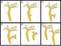
1. DNA
2. Enhancer
3. Promoter
4. Gene
5. Transcription Activator Protein
6. Mediator Protein
7. RNA Polymerase
- Past Related Projects: Wikipedia:WikiProject AP Biology Bapst 2012, Wikipedia:WikiProject AP Biology Bapst 2013, Wikipedia:WikiProject AP Biology Bapst 2014, Wikipedia:WikiProject AP Biology Bapst 2015, Wikipedia:WikiProject AP Biology 2016, & Wikipedia:WikiProject AP Biology 2017
A high school class in Maine - will contribute images to Wikipedia article and the commons until June 10, 2017. The collective goal is to contribute excellent biology diagrams to the Commons and to corresponding Wikipedia articles. This is done as part of an Advanced Placement Biology course. The lead editor is Chris Packard. This project is inspired by the 2009 Wikipedia AP Biology Project. There are many basic and important diagrams missing from biological articles and we're doing our part to fix this.
- Students will work alone, there are 49 students so we should have 49 new images with captions and labels.
- The time frame will be three weeks.
- Students will be required to write a summary of why they select a topic; hopefully, eliminating obscure, random topic selections. They also must create labels and captions for their photos
- They may add it to encyclopedia articles.
- The best of the bunch will be submitted as Wikipedia featured pictures, see other candidates here. Featured images must be in .svg (vector) format.
Feel free to discuss this project. Please notify me of any concerns; especially if they involve the behavior of my students on Wikipedia. With a little patience, this should be an inspirational experience for all.
Goals / Motivation
- To create a situation that not only vigorously enhances our ability to make quality decisions but also to improve our traction on the roads of 'Merica
- To improve the images in Wikipedia's coverage of Biology articles.
- To encourage promising students to write, create, learn, and contribute volunteer efforts through a service learning project.
- The dreaded “Research Project” is a standard hurdle for most AP Programs. Rightfully so, being that many college courses require such publications to validate your existence. This new approach to constructing a scientific document, is far more authentic and interesting. Rather than researching for a paper that is destined for the teacher's eyes and then a one way trip to the circular bin, let us contribute to the world-wide data base for others to benefit. I hope this will be an interesting and memorable project and assessment. It's funny, I can remember a number of projects and papers I wrote during my own high school experience, but I can remember no tests whatsoever.
Contributions
As you upload your projects and add them to Wikipedia please add them to the gallery below. By adding a new line which begins with the word "File" and them follows the format of my sample image. Make sure to include your caption.
-
 The aortic valve controls outflow of blood from the left ventricle of the heart through the aorta (valve is indicated within the yellow highlighted box). Normal aortic valve is tricuspid. Five types of bicuspid valve are shown, with Type 1 being most prevalent. Bicuspid valve forms when the tissue surrounding one of the cusps (leaflets) of the valve fuse during fetal development. This developmental anomaly can have either negative or no effect on the individual.
 -
 The process and possible outcomes of random X chromosome inactivation in female human embryonic cells undergoing mitosis. 1.Early stage embryonic cell of a female human 2.Maternal X chromosome 3.Paternal X chromosome 4.Mitosis and random X chromosome inactivation event 5.Paternal chromosome is randomly inactivated in one daughter cell, maternal chromosome is inactivated in the other 6.Paternal chromosome is randomly inactivated in both daughter cells 7.Maternal chromosome is randomly inactivated in both daughter cells 8.Three possible random combination outcomes
 -
 This pedigree has a sex-linked recessive disorder. It is possible to tell the trait is recessive because it skips a generation with only carriers in generation 2. Because the trait is sex-linked only females can be carriers since only females have 2 x chromosomes. As a result a lot more males tend to display sex-linked recessive disorder than females. Female carriers tend to give the trait to about half of their sons and daughters but only the sons will be affected while the daughters will only be carriers.
 -
 This pedigree has a sex-linked dominant disorder. It is possible to tell it is dominant because it doesn't skip generations. It is possible to tell it is sex-linked because affected fathers will pass the trait on to all of their daughters. This is because they have to pass along their affected dominant x but none of their sons because they pass on a y to their sons. The affected mother in the second generation is heterozygous (having a dominant and a recessive allele) will have a 50% chance of passing the trait on to all of her children regardless of their gender.
 -
 This pedigree has a autosomal recessive disorder. It is possible to tell this order is recessive because it skips generations with none of generation 3 actually displaying the trait. When both parents display the disorder all of their kids have to display the disorder. But when only one parent displays the disorder all of the kids will be carriers but unaffected by the trait. It is possible to tell the trait is autosomal because it affects females and males equally.
 -
 This pedigree has an autosomal dominant disorder. It is possible to tell the trait is dominant because it never skips a generation and there are no unaffected carriers. Affected children must have affected parents. When one parent possesses the trait and is heterozygous ( possessing a dominant and recessive allele) approximately half of the children will possess the dominant disorder.
 -
 The left side of this diagram shows the plant pathway of this relationship, where the host plant transfers between 4% to 20% of its photosynthetically fixed carbon to the mycorrhiza. On the right side of this diagram, the arbuscular mycorrhiza pathway, which branches off from the plant root, provides the plant with nutrients, including, most importantly, phosphate and nitrogen.
 -
 In both stages of metamorphosis, the insect begins the cycle as an egg. In a complete metamorphosis the insect passes through four distinct phases which produce an adult that does not resemble the larvae. In an incomplete metamorphosis an insect does not go through a full transformation, but instead transitions from a nymph to an adult by molting its exoskeleton.
 -
 This image shows a priming web built from different types of priming. The lines in this web indicate associations that an individual might have. If two words are more closely linked in the web, then they are more likely to be more quickly recognized when primed with a nearby word. The dotted lines indicate morpheme primes, or primes from words that sound similar to each other, while the straight lines indicate semantic primes or words that have meanings or associations that relate to each other.
 -
 Diagram 1- Testosterone, shown in this image as T, outside of a cell containing androgen receptors and 5a-reductase (5a-r), an enzyme which converts T to DHT. AR stands for androgen receptor, which binds to both T and DHT. Diagram 2 - Testosterone (T) inside the cell, going one of two paths.(1) T does not bind to the enzyme 5a-reductase (5a-R) and simply binds to the androgen receptor (AR), where it will later be transported into the nucleus.(2) T binds with the enzyme 5a-R, becoming DHT (indicated by the descending arrow). From there, it binds to the AR and will be transported to the nucleus.
 -
 Predator-prey coevolution causes favorable changes in both species. In this example, owls cause changes in mice and vice-versa. Light mice are easily hunted by all owls, so they don't survive to maturity. Therefore, they cannot pass down their genes, so their population declines. Since dark mice are harder to see, they are able to survive and pass on their genes. Owls with poor eyesight cannot see the dark mice, so they starve and their population declines. Since owls with good eyesight can effectively hunt dark mice they can eat and reproduce, passing down their genes. After some time the mouse and owl populations have changed from their original state.
 -
 Key:
Key:
(a) Sodium (Na+) ion
(b) Potassium (K+) ion
(c) Sodium channel
(d) Potassium channel
(e) Sodium-Potassium Pump
In the stages of an action potential, the permeability of the membrane of the neuron changes. At the resting state (1), sodium and potassium ions are unable to pass through the membrane, and the neuron has a negative charge inside (mainly due to the large proteins that are negatively charged, as well as the lower amount of positive K+ ions inside the neuron). Once the action potential is triggered, the depolarization (2) of the neuron activates the sodium channel, allowing sodium ions to pass through the membrane of the neuron and results in a positive charge in the neuron and a negative charge in the extracellular fluid. After the action potential is reached, the neuron begins repolarization (3), where the sodium channels close and the potassium channels open, allowing potassium ions to cross the membrane and flood into the extracellular fluid, resulting in a positive charge in the extracellular fluid and a negative charge that is below the resting potential of the neuron. Finally, to return the neuron to that resting potential after the potassium pump closes, a sodium-potassium pump works to exchange three sodium ions per two potassium ions across the plasma membrane during the refractory period (4). Once the Na+ and K+ are back where they started, the neuron is back to its resting state (1), ready to do it all over again for the next action potential. -
 This image shows the difference between endotherms and ectotherms. The mouse is endothermic and regulates its body temperature through homeostasis. The lizard is ectothermic and its body temperature is dependent on the environment
 -
 This is a diagram showing changes in genetic sequences, called mutations. Figure (1) shows a normal nucleotide sequence, consisting of 4 codons with 3 base pairs. Figure (2) shows a missense mutation, where the base pair cytosine has been changed to guanine. This does not change other base pairs in the codon. This one has the least effect on the gene, because only one amino acid will change. Figure (3) shows a deletion, where one base pair is deleted from the sequence. This causes a shift of all the base pairs after the deletion point. Figure (4) shows an insertion in the third base pair of the third codon. This causes all of the proceeding base pairs to be shifted one to the right, causing changes in a gene sequence. Figure (5) shows a frameshift mutation. This is when the codons shift over one position, causing differences in amino acid sequences. Figure (6) is a repeat expansion. This is when an entire codon replicates itself one or more times. This causes a repeat in amino acid sequences, causing proceeding codons to shift over one or more positions.
 -
 This diagram shows the relationship between different families of turtles within the superfamily of Testudinoidea. The four primary families within the superfamily of Testudinoidea are Emydidae, Geoemydidae, Platysternidae, and Testudinae. The family of Emydidae is further subdivided into the families of Dierochelyinae and Emydinae. The family of Geoemydidae is further subdivided into Geoemydinae and Rhinoclemmydinae.
 -
 This image shows two types of root systems in plants that provide their stems and leaves with water and mineral. The fibrous root system is characterized by many roots with similar sizes. In contrast, plants that use the taproot system grow a main root with smaller roots branching off of the taproot. Where the letters start mark the beginning of the roots. A. Fibrous Root System B. Taproot System
 -
 An example of membrane receptors. (1.) Ligands, located outside the cell. (2.) Ligands connect to specific receptor proteins based on the shape of the active site of the protein. (3.) The receptor releases a messenger once the ligand has connected to the receptor.
 -
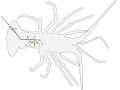 The figure above displays the reflex arc of a lobster in order to activate the caridoid escape reaction. (A) Microscopic hairs etched along the tail of the decapod activate a somatic signal in response to the presence of an environmental stimulus. (B) The action potential activated by the somatic interneuron relays an impulse to the lateral giant (LG) interneuron. (C) The lateral giant interneuron executes a reflex by relaying impulses to various giant motor neurons within the abdomen of the lobster. The muscular contractions result in the lobster being able to successfully propel itself through the water, away from the site of stimulus.
 -
 1) Antibodies (A) and pathogens (B) free roam in the blood. 2) The antibodies bind to pathogens, and can do so in different formations such as: opsonization (2a), neutralisation (2b), and agglutination (2c). 3) Phagocytosis begins when a phagocyte (C) approaches the pathogen. The Fc region (D) of the antibody binds to one of the Fc receptors (E) on the phagocyte. 4) Phagocytosis occurs as the pathogen is ingested.
 -
 -Step 1- Water imbibition, the uptake of water, results in rupture of seed coat. -Step 2-The imbibition of the seed coat results in emergence of the radicle (1) and the plumule(2), the cotyledons get unfolded(3). -Step 3-This marks the final step in the germination of the seed where the cotyledons are expanded which are the true leaves/peasNote- Temperature must be kept at an optimum level.
 -
 The humoral immune response occurring after a pathogen invades the body.
 -
 Lysogenic Cycle:1. The prokaryotic cell is shown with its DNA, in green. 2. The bacteriophage attaches and releases its DNA, shown in red, into the prokaryotic cell. 3. The phage DNA then moves through the cell to the host’s DNA. 4. The phage DNA integrates itself into the host cell's DNA, creating prophage. 5. The prophage then remains dormant until the host cell divides. 6. After the host cell has duplicated, the phage DNA in the the daughter cells activate, and the phage DNA begins to express itself. Some of the cells containing the prophage go on to create new phages which will move on to infect other cells.
 -
 This is a diagram of a normal ovary going through it’s cycle, with a polycystic ovary showing how the cycle messes up and forms cysts.
 -
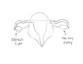 This is a diagram of how an ovary looks with a cyst.
 -
 This diagram shows six stages of how the hydra organism asexually reproduces by the process of budding.1. This frame pictures a normal hydra organism.2. The hydra has begin to bud, creating a bump on itself, because it is using its DNA and outer-body membrane to create an identical copy to itself.3. The daughter hydra has begun to grow out from the parent hydra.4. The daughter continues to grow larger from the parent hydra, and the connecting membrane between the two organisms has begun to cleave from each other.5. The daughter hydra has broken off from its parent organism to create two different entities.6. The daughter hydra has grown to become exactly identical to its parent hydra due to the same DNA from the budding process, and they are both ready to asexually reproduce again.


.svg)


.svg)
.svg)


.svg)

.svg)
.svg)


Contributors
Add your user name here following my example. Just add this template with your username instead of the line: {{user|username}} and then, if your username is not identifiable, your real first name.
- Earthdirt (talk · contribs) - Chris (AKA Mr. Packard) - IMAGE TOPIC NAME HERE
- noahrob (talk · contribs) - Noah - Priming Web Diagram
- lcaron101 (talk · contribs) - Lydia - Bicuspid Aortic Valve
- Lexicunningham1 (talk · contribs) - Lexi - Negative feedback
- F-150_Smiley (talk · contribs) - Ford - Turtle Cladogram
- Maher33 (talk · contribs) - Maher - Opsonization
- LFrancis19 (talk · contribs) - Landyn - Immune system
- Maddieahola (talk · contribs) - Maddie - Gravitropism
- reh0318 (talk · contribs) - Rachel - Humoral Response
- Tessa has dibs on the Leaf thing
- {{user|Hydra2114 - Catherine - Pedigree
- Sseifert242 (talk · contribs) - Sarah - Lysogenic cycle
- sxs5ux7 (talk · contribs) - Shay - Ligase
- Ebagley18 (talk · contribs) - Ethan - Dead zone
- CThompson20 (talk · contribs) - Claire - Action Potential permeability of membrane
- ussypme18 (talk · contribs) - Caroline - Mycorrhizae
- ccaldwell19 (talk · contribs) - Colby - Natural selection
- Gretafrost (talk · contribs) - Greta - Hydrangea flower color and soil pH
- A1yssa18 (talk · contribs) - Alyssa - Alzheimer brain and normal brain comparison
- jenna.leighton (talk · contribs) - Jenna - Ovarian disease
- Windover14 (talk · contribs) - Lucas - Duck feet counter current exchange
- lilymclaughlin01 (talk · contribs) - Lily- X-inactivation
- ElinorHunt (talk · contribs) - Ellie - Ischemic stroke
- Noahjgagne (talk · contribs) - Noah- Seed Germination
- User-multi error: no username detected (help). - Devin- Pressure Flow Hypothesis or transpiration
- A.houghton19 (talk · contribs) - Abby - budding
- wpyzynski (talk · contribs) - Wyatt - Receptor (biochemistry)
- User-multi error: no username detected (help). - Kelena - Metapopulation
- XXx_m1n10n5_xXx (talk · contribs) - Will H - Reflex arc in Decapoda with the LG interneuron
- KaitlinLiu (talk · contribs) - Kaitlin - Taproot & Fibrous Root System
- VictorTheWhite (talk · contribs) - Victor - Testosterone bonding
- bigman24225 (talk · contribs) - Noah Missbrenner - Interspecific Competition
- Ijeoma__Obi (talk · contribs) - Ijeoma Obi - Anaerobic respiration
- grcoffey (talk · contribs) - Gabe Coffey EPO Effect on Athletic Performance in Distance Runners
- reyasingh56 (talk · contribs) - Reya Singh - Immunotherapy
- mbrookings19 (talk · contribs) - Maddie Brookings - Lake Stratification
- Turbotronbabyjesus9000 (talk · contribs) - Sam - Binary Fission
- User-multi error: no username detected (help). - Elliot - Positive Feedback
- apbio2018 (talk · contribs) - Lily C - Disinfectant
- gnomstah (talk · contribs) - Naomi Moynihan - Smallpox Vaccine
- edesjardins19 (talk · contribs) - Erica Desjardins - Visual Impairment
- PlasticH8r (talk · contribs) - Alex - Plastiglomerate
- jordan_hawes (talk · contribs) - Jordan Hawes - Endotherm
- Liamdunn2000 (talk · contribs) - Liam Dunn - Mutations
- Noahjgagne (talk · contribs) - Noah Gagne Seed Germination
- Williamtobin173682 (talk · contribs) - William Tobin Phage Therapy
- KyleJeffrey (talk · contribs) - Kyle Jeffrey Cas9
Uploading
In order to complete the assignment and reap all the benefits of your hard work (such as a good grade) you MUST complete all of the following steps. If you need help, just ask.
How to, step by step
Step 1: Create a Wikipedia Global account by clicking "Login/create account" in the upper right hand corner of this page.
Step 2: Click here to use the WikiCommons File Upload Wizard
Step 3: If you didn't do it in the Wizard, categorize your image by adding a one or more [[Category:_______]] tags at the bottom of the page (fill in the name of the category in the _______.) You might use Category:Biology diagrams (but that's not a very helpful category) or something more specific like Category:Molecular biology or something else appropriate.
Step 4: If you didn't do it in the Wizard you should also now add your labels and your caption information in the description to your upload page in the Commons.
Step 5: Your image is now available in all Wiki Projects, including Wikipedia. So let's add it to the article! Go to the article you want to add your donated image to. In the top of the section of the article or the subheading you want to add the image to add something like this:
[[File:MY IMAGE NAME.png|right|thumb|200px|The [[caption]] of '''my image'''.]]
That's not too hard is it? For your caption you'll need to follow Wikipedia style and use some mark up to do this - it's kind of like a micro-essay. The [[ ]] creates a link to the given page on Wikipedia and the ''' ''' make the word bold, in Wikipedia it's appropriate to bold the title of the article the first time it's used in the text or in a caption."
Step 6: Wow you've done it! Now you just have to turn in your work by adding it to gallery in the section above here called "Contributions". Just follow the model I provided in the first entry. Make sure that your entry is between the <gallery> and </gallery> tags or it won't show up. Your caption will likely have to be shorter than your description, see the style advice below.
Style guides
To get past the stumbling blocks of editing Wikipedia, articles will have to conform to the Wikipedia style guides. The largest barriers are:
- Wikipedia:Manual of Style/Images - The basic overview of images (the Wikipedia:Picture tutorial is also useful.
- Wikipedia:Manual of Style/Captions - Writing a good caption may be harder than you think.
- Wikipedia:Copyrights - Make sure to post a license on your image which releases all copyrights and makes it free use image AND don't use images from anywhere except the Commons if your image integrates other images.
- Wikipedia:File names - Pick the right name for your file.
- Wikipedia:Preparing images for upload - Pick the right file type (images created using entirely Google Draw should be saved as .SVG, whereas most other images you make will be saved as a .PNG in rare cases an a .JPG or .JPEG can be used)
- Wikipedia:Uploading images or WikiCommons Uploading Images - Do it right the first time (or just use the Wizard).
- Wikipedia:Ten things you may not know about images on Wikipedia - Kind of interesting.
You can always ask for help at:
Writing a good image caption
There are several criteria for a good caption. A good caption:
- clearly identifies the subject of the picture, without detailing the obvious.
- is succinct (that means short).
- establishes the picture's relevance to the article.
- provides context for the picture.
- draws the reader into the article.
Different people read articles different ways. Some people start at the top and read each word until the end. Others read the first paragraph and scan through for other interesting information, looking especially at pictures and captions. For those readers, even if the information is adjacent in the text, they will not find it unless it is in the caption—but do not tell the whole story in the caption—use the caption to make the reader curious about the subject.
Another way of approaching the job: imagine you're giving a lecture based on the encyclopedia article, and you are using the image to illustrate the lecture. What would you say while attention is on the image? What do you want your audience to notice in the image, and why? Corollary: if you have got nothing to say, then the image probably does not belong in the article.
Images for the lead
It is very common to use an appropriate representative image for the lead of an article, often as part of an infobox. The image helps to provide a visual association for the topic, and allows readers to quickly assess if they have arrived at the right page. For most topics, the selection of a lead image is plainly obvious: a photograph or artistic work of a person, photographs of a city, or a cover of a book or album, to name a few.
Image selection for other topics may be more difficult and several possible choices could be made. While Wikipedia is not censored, as outlined in the above section on offensive images, the selection of the lead image should be made with some care with respect to this advice. Lead images are loaded and shown upon navigating to the page, and are one of the first things that readers will see. Editors should avoid using images that readers would not have expected to see when navigating to the page. Unlike other content on a page that falls below the lead, the lead image should be chosen with these considerations in mind.
Some advice on selecting a lead image include the following:
- Lead images should be images that are natural and appropriate visual representations of the topic; they not only should be illustrating the topic specifically, but should also be the type of image that is used for similar purposes in high-quality reference works, and therefore what our readers will expect to see. Lead images are not required, and not having a lead image may be the best solution if there is no easy representation of the topic.
- Lead images should be selected to be of least shock value; if an alternative image exists that still is an accurate representation of the topic but without shock value, it should always be preferred. For example, using an image of deportees being subjected to selection as the lead image at this version of Holocaust is far preferable to the appropriate images that appear later in the article that show the treatment of the prisoners or corpses from the camps.
- Sometimes it is impossible to avoid the use of a lead image with perceived shock value if the topic itself is of that nature, for example in articles on various parts of human genitalia. It should be anticipated, through Wikipedia:Content disclaimer, that readers will be aware they will be exposed to potentially shocking images when navigating to articles on such topics.
Planning and resources
- Wikipedia tutorials for beginners
- Editing commands cheatsheet
- Getting started
- The perfect article
- Assessment
- Article development
- Peer Review
- [[Active gif creator]]
Talk pages
These are places where you can leave and receive messages and questions, every page has one. Whenever you edit these pages, make sure that you are signed in. Also, add four tildes ~~~~ to the end of all comments you make on talk pages. This will let people know who is talking.