
Corpus cavernosum penis: Difference between revisions
Genglonghsu (talk | contribs) |
|||
| Line 38: | Line 38: | ||
Image:Illu penis.jpg|Structure of the penis |
Image:Illu penis.jpg|Structure of the penis |
||
Image:Gray543.png|The deeper branches of the [[internal pudendal artery]]. |
Image:Gray543.png|The deeper branches of the [[internal pudendal artery]]. |
||
Image: |
Image:Penile anatomy cross.jpg|The penis in transverse section, showing the bloodvessels. |
||
Image:Gray1136.png|Male pelvic organs seen from right side. |
Image:Gray1136.png|Male pelvic organs seen from right side. |
||
Image:Penile anatomy side.jpg|Diagram of the arteries and the veins of the [[penis]]. |
Image:Penile anatomy side.jpg|Diagram of the arteries and the veins of the [[penis]]. |
||
Revision as of 13:26, 8 April 2019
This article needs additional citations for verification. (September 2009) |
| Corpus cavernosum penis | |
|---|---|
| File:Penile anatomy cross.jpg Transverse section of the penis. | |
 The constituent cavernous cylinders of the penis. | |
| Details | |
| Identifiers | |
| Latin | corpus cavernosum penis |
| TA98 | A09.4.01.014 |
| TA2 | 3678 |
| FMA | 19618 |
| Anatomical terminology | |
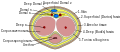
A corpus cavernosum penis (singular) (cavernous body of the penis) is one of a pair of sponge-like regions of erectile tissue, the corpora cavernosa (plural) (cavernous bodies), which contain most of the blood in the penis during an erection.[1][2][3] Such a corpus is homologous to the corpus cavernosum clitoridis in the female; the body of the clitoris contains erectile tissue in a pair of corpora cavernosa (literally "cave-like bodies") with a recognisably similar structure.
Anatomy
The two corpora cavernosa and corpus spongiosum (also known as the corpus cavernosum urethrae in older texts and in the adjacent diagram) are three expandable erectile tissues along the length of the penis, which fill with blood during penile erection. The two corpora cavernosa lie along the penis shaft, from the pubic bones to the head of the penis, where they join. These formations are made of a sponge-like tissue containing trabeculae, irregular blood-filled spaces lined by endotheliumand separated by an incomplete septum-connective tissue septa which is dorsally fenestrated.connective tissue septa.[4][5] [6]
The male anatomy has no vestibular bulbs, but instead a corpus spongiosum, a smaller region along the bottom of the penis, which contains the urethra and traverses distally to the glans penis.[6]
Physiology
In some circumstances, a release of nitric oxide precedes relaxation of muscles in the corpora cavernosa and corpus spongiosum, in a process similar to female arousal. The spongy tissue fills with blood, from arteries down the length of the penis. A little blood enters the corpus spongiosum; the remainder engorges the corpora cavernosa, which expand to hold 90% of the blood involved in an erection, increasing both in length and in diameter. The function of the corpus spongiosum is to prevent compression of the urethra whereby ejaculation patency is secured by a distal ligament-os analog particularly in intravaginal coitus, during erection.
Blood can leave the erectile tissue only through a drainage system of veins (most importantly, one Deep Dorsal Vein, a pair of cavernosal veins, and two pairs of para-arterial veins, which are collectively coined "the erection-related veins", between the Buck's fascia and the tunica albuginea), away from the corpus cavernosum via emissary veins. The new understanding of the erection-related veins differs from just one single deep dorsal vein in the older text [5] [6] [7] The expanding spongy tissue presses against the outer longitudinal layer of the surrounding dense tissue (tunica albuginea) constricting these veins, preventing blood from leaving. Consequently, the penis becomes rigid as a result. The glans penis, buttress with a distal ligament-os analog which is an integral structure of the corpora cavernosa, remains more malleable during erection because its perisinusoidal shell is full of fibro-elastic components which is much more than elsewhere in the penis.[8]
Additional images
-
 Structure of the penis
 -
 The deeper branches of the internal pudendal artery.
 -
The penis in transverse section, showing the bloodvessels.
-
 Male pelvic organs seen from right side.
 -
Diagram of the arteries and the veins of the penis.
-
 Diagram of the arteries of the penis.
 -
 Cross section of penis.
 -
 Medical ultrasonography of a normal penis.


References
- ^ Werner Lierse (6 December 2012). Applied Anatomy of the Pelvis. Springer Science & Business Media. ISBN 978-3-642-71368-2.
- ^ Heide Schatten; Gheorghe M. Constantinescu (21 March 2008). Comparative Reproductive Biology. John Wiley & Sons. ISBN 978-0-470-39025-2.
- ^ Michele Bertolotto (22 December 2007). Color Doppler US of the Penis. Springer Science & Business Media. pp. 157–. ISBN 978-3-540-36677-5.
- ^ "Embarrassing erections". 2007-01-17. Archived from the original on 2007-01-02. Retrieved 2007-01-17.
{{cite web}}: Unknown parameter|deadurl=ignored (|url-status=suggested) (help) - ^ "Understanding a man's erection". whitelotuseast.[better source needed]
- ^ a b c "Penis Structure". Academic Press.
- ^ "Penis Structure-Erection". Academic Press.
- ^ "Erection Abnormality". Academic Press.
External links
- Anatomy photo:42:st-1102 at the SUNY Downstate Medical Center - "The Male Perineum and the Penis: Penis"