
Colic flexures
| Colic flexures | |
|---|---|
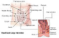
 Colorectal anatomy (the splenic flexure is labeled at upper right, the hepatic flexure at upper left) | |
 | |
| Details | |
| Precursor | Hindgut |
| Identifiers | |
| Latin | Flexura coli |
| FMA | 14555 |
| Anatomical terminology | |
There are two colic flexures, or curvatures in the transverse colon. The one on the right, the right colic flexure is known as the hepatic flexure.
The one on the left, the left colic flexure is known as the splenic flexure.
Structure
The right colic flexure or hepatic flexure (as it is next to the liver) is the sharp bend between the ascending colon and the transverse colon. The hepatic flexure lies in the right upper quadrant of the human abdomen. It receives blood supply from the superior mesenteric artery.
The left colic flexure or splenic flexure (as it is close to the spleen) is the sharp bend between the transverse colon and the descending colon. The splenic flexure is a watershed region as it receives dual blood supply from the terminal branches of the superior mesenteric artery and the inferior mesenteric artery, thus making it prone to ischemic damage in cases of low blood pressure because it does not have its own primary source of blood. In the context of ischemia, the splenic flexure is sometimes referred to as Griffith's point, along with the upper rectum (Sudak's point).
See also
Additional images
-
 Intestines
 -
 The duodenum and pancreas.
 -
 Double Contrast Barium Enema - Using Positive and Negative Contrast


External links
- Lotti M. Anatomy in relation to left colectomy
- Anatomy photo:37:13-0102 at the SUNY Downstate Medical Center
- Anatomy image:8182 at the SUNY Downstate Medical Center
This human digestive system article is a stub. You can help Wikipedia by expanding it. |