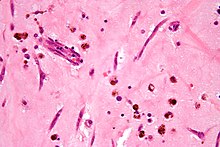
Myxoid tumor
Appearance
A myxoid tumor is a connective tissue tumor with a "myxoid" background, composed of clear, mucoid substance.[1]

This tumoral phenotype is shared by many tumoral entities:
- Myxomas
- Myxoid hamartoma
- Aggressive angiomyxoma
- Myxoid leiomyoma
- Chondromyxoid fibroma
- Myxoid neurofibroma
- Nerve sheath myxoma (neurothekeoma)
- Myxolipoma
- Angiomyofibroblastoma
- Myxoid leiomyosarcoma
- Myxoid liposarcoma
- Lipoblastoma
- Myxofibrosarcoma
- Myxoid cortical adenoma
- Pleomorphic adenoma
- Undifferentiated embryonal sarcoma
- Plexiform angiomyxoid myofibroblastic tumor
- Myxoid plexiform fibrohistiocytic tumor
- Angiomyxolipoma (vascular myxolipoma)
- Parachordoma
- Acral myxoinflammatory fibroblastic sarcoma
References
- ^ Willems, S. M.; Wiweger, M; Van Roggen, J. F.; Hogendoorn, P. C. (2010). "Running GAGs: Myxoid matrix in tumor pathology revisited: What's in it for the pathologist?". Virchows Archiv. 456 (2): 181–92. doi:10.1007/s00428-009-0822-y. PMC 2828560. PMID 19705152.
