Blastocoel
This section needs expansion with: at least the further information to make understandable the content of the images that appear in the infobox. You can help by adding to it. (January 2016) |
| Blastocoel | |
|---|---|
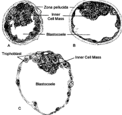
 Descriptive figure legend goes here. Panel A is… Panel B is… Panel C is… [citation needed] | |
 Schematic diagram showing the blastocyst, with its embryoblast (inner cell mass) and its trophoblast layer, alongside the surface of the endometrium.[citation needed] | |
| Details | |
| Carnegie stage | 3 |
| Days | 5 |
| Precursor | morula[citation needed] |
| Gives rise to | gastrula,[citation needed] primitive yolk sac[citation needed] |
| Anatomical terminology | |
A blastocoel (alt. spelling blastocoele, blastocele),[1] also termed the blastocyst cavity[2][better source needed] (or cleavage or segmentation cavity[1]) is the name given to the fluid-filled cavity of the blastula (blastocyst) that results from cleavage of the oocyte (ovum) after fertilization.[1][2] It forms during embryogenesis,[citation needed] as what has been termed a "Third Stage" after the single-celled fertilized oocyte (zygote, ovum[citation needed]) has divided into 16-32 cells,[2] via the process of mitosis.[citation needed] It can be described as the first cell cavity formed as the embryo enlarges,[citation needed] the essential precursor for the differentiated, topologically distinct, gastrula.[citation needed] The adjectival form of blastocoel is blastocoelic.[1][3][4][5]
A blastocoel is a fluid-filled cavity that forms in the animal hemisphere of early amphibian and echinoderm embryos, or between the epiblast and hypoblast of avian, reptilian, and mammalian blastoderm-stage embryos.
Mammalian blastocoel
After fertilization, the mammalian cells, called blastomeres, undergo rotational cleavage until they are at the 16-cell stage called the morula. The morula has a small group of internal cells surrounded by a larger group of external cells. These internal cells are called the inner cell mass (ICM) and will go on to become the actual embryo. The external, surrounding cells develop into the trophoblast cells. However, at this stage there is no cavity within the morula; the embryo is still a ball of dividing cells. In a process called cavitation, the trophoblast cells secrete fluid into the morula to create a blastocoel, the fluid-filled cavity. The membranes of the trophoblast cells contain sodium (Na+) pumps, Na+/K+- ATPase and Na+/H+ exchangers, that pump sodium into the centrally forming cavity. The accumulation of sodium pulls in water osmotically, creating and enlarging the blastocoel within the mammalian embryo (Borland 1977; Ekkert et al. 2004; Kawagishi et al. 2004). The oviduct cells stimulate these trophoblast sodium pumps as the fertilized egg travels down the fallopian tube towards the uterus (Xu et a. 2004). As the embryo further divides, the blastocoel expands and the inner cell mass is positioned on one side of the trophoblast cells forming a mammalian blastula, called a blastocyst.
References
Borland R M. Transport processes in the mammalian blastocyst. Dev. Mammals. 1977;1:31–67.
Wiley L M. Cavitation in the mouse preimplantation embryo: Na/K ATPase and the origin of nascent blastocoel fluid. Dev. Biol. 1984;105:330–342.
- ^ a b c d Dorlands Staff (2004). "blastocoele [dictionary entry]". Dorland's Illustrated Medical Dictionary (online). Amsterdam, NDE: Elsevier-Saunders. Retrieved 30 January 2016.
blastocoele… [blasto- + -coele] the fluid-filled cavity of the mass of cells (blastula) produced by cleavage of a fertilized ovum. Sometimes spelled… [c]alled… [Also] blastocoelic… pertaining to the blastocoele.
- ^ a b c Senn, A.; Schöni-Affolter, F.; Dubuis-Grieder, C. & Strauch, E.; et al. (2007). "Module 8, Embryonic Phase; [Section] 8.1 The Carnegie Stages; Synoptic Table of the Carnegie Stages 1 - 6; [page] 'Stage 3, Approx. 4th - 5th day, 0.1 - 0.2 mm' [rev. 25.04.07]". In Manuèle Adé-Damilano (ed.). embryology.ch: Online Course in Embryology for Medicine Students. Fribourg, CHE: Swiss Virtual Campus, l'Université de Fribourg, et al. Retrieved 30 January 2016.
Drs Senn and Dubuis-Grieder are at l'Université Lausanne, Drs Adé-Damilano and Schöni-Affolter at l'Université de Fribourg, and Dr Strauch at Universität Bern.
{{cite book}}: CS1 maint: multiple names: authors list (link) - ^ Ereskovsky, Alexander V. (2010). The Comparative Embryology of Sponges. Springer. ISBN 978-90-481-8574-0.
- ^ Mader S. S. (2000): Human biology. McGraw-Hill, New York, ISBN 0-07-290584-0; ISBN 0-07-117940-2.
- ^ Gilbert S. F. (2010). Developmental Biology (Ninth ed.). Sinauer Associates. ISBN 978-0-87893-558-1.
