
Great saphenous vein
This article needs additional citations for verification. (November 2013) |
| Great saphenous vein | |
|---|---|
 The great saphenous vein and landmarks along its course | |
 The great saphenous vein and its tributaries at the fossa ovalis in the groin. | |
| Details | |
| Source | dorsal venous arch of the foot, and others |
| Drains to | femoral vein |
| Identifiers | |
| Latin | vena saphena magna |
| TA98 | A12.3.11.003 |
| TA2 | 5058 |
| FMA | 21376 |
| Anatomical terminology | |
The great saphenous vein (GSV), previously also called the long saphenous vein, is a large, subcutaneous, superficial vein of the leg. It is the longest vein in the body running along the length of the lower limb.
The terms "saphaina" (Greek, meaning "manifest," "to be clearly seen") and "safoon" (Hebrew, "שָׂפוּן" meaning "hidden/covered") as well as "safin" (Arabic, "سافن" meaning "deep/embedded")[1] have been claimed as the origin for the word "saphenous."[1]
Structure
The great saphenous vein originates from where the dorsal vein of the big toe (the Hallux) merges with the dorsal venous arch of the foot. After passing in front of the medial malleolus (where it often can be visualized and palpated), it runs up the medial side of the leg. At the knee, it runs over the posterior border of the medial epicondyle of the femur bone. The GSV then courses anteriorly to lie on the anterior surface of the thigh before entering an opening in the fascia lata called the saphenous opening. It forms an arch, the saphenous arch, to join the common femoral vein in the region of the femoral triangle at the sapheno-femoral junction. [citation needed]
Tributaries
At the ankle it receives branches from the sole of the foot through the medial marginal vein; in the lower leg it anastomoses freely with the small saphenous vein, communicates by perforator veins (Cockett perforators) with the anterior and posterior tibial veins and receives many cutaneous veins; near the knee it communicates with the popliteal vein by the Boyd perforator, in the thigh it communicates with the femoral vein by perforator veins (Dodd perforator) and receives numerous tributaries; those from the medial and posterior parts of the thigh frequently unite to form a large accessory saphenous vein which joins the main vein near the sapheno-femoral junction.[2]
Near the fossa ovalis it is joined by the superficial epigastric, superficial iliac circumflex, and superficial external pudendal veins.
The thoracoepigastric vein runs along the lateral aspect of the trunk between the superficial epigastric vein below and the lateral thoracic vein above and establishes an important communication between the femoral vein and the axillary vein.
Clinical significance
Pathology of the great saphenous vein is relatively common, but in isolation typically not life-threatening.[3]
- Varicose veins: The great saphenous vein, like other superficial veins, can become varicose; swollen, twisted and lengthened, and generally considered to be unsightly. Varicose veins are not life-threatening and various treatment options are available.
- Thrombophlebitis: The GSV can thrombose. This type of phlebitis of the GSV is usually not life-threatening in isolation; however, if the blood clot is located near the sapheno-femoral junction or near a perforator vein, a clot fragment can migrate to the deep venous system and to the pulmonary circulation. Also it can be associated with, or progress to a deep vein thrombosis which must be treated promptly. So a GSV thrombosis is investigated by ultrasonography to detect if these complications are present.[3]
Use in cardiovascular procedures
The vein is often removed by cardiac surgeons and used for autotransplantation in coronary artery bypass operations, when arterial grafts are not available or many grafts are required, such as in a triple bypass or quadruple bypass.
The great saphenous vein is the conduit of choice for vascular surgeons,[4][5] when available, for doing peripheral arterial bypass operations [ see vascular bypass ] because it has superior long-term patency compared to synthetic grafts (PTFE, PETE (Dacron)), human umbilical vein grafts or biosynthetic grafts [Omniflow]. Often, it is used in situ (in place), after tying off smaller tributaries and destruction of the venous valves with a device called valvulotome, e.g. LeMaitre's valvulotome.
Removal of the saphenous vein will not hinder normal circulation in the leg. The blood that previously flowed through the saphenous vein will change its course of travel. This is known as collateral circulation.
The saphenous nerve is a branch of the femoral nerve that runs with the great saphenous vein and can be damaged in surgery on the vein.
Use in emergency medicine
When emergency resuscitation with fluids is necessary, and standard intravenous access cannot be achieved due to venous collapse, saphenous vein cutdown may be necessary.
See also
Additional Images
-
 Superficial veins oflower limbSuperficial dissection. Anterior view.
 -
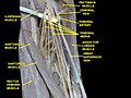 GREAT SAPHENOUS VEIN. Deep dissection. Anterior view.
 -
 Illustration depicting veins of the leg including great saphenous vein (anterior view).


References
- ^ a b Caggiati, Alberto; Bergan, John J. (2002). "The saphenous vein: derivation of its name and its relevant anatomy". Journal of Vascular Surgery. 35 (1): 172–5. doi:10.1067/mva.2002.118826. PMID 11802151.
- ^ Franceschi, C.; Zamboni, P. (2009). Principles of Venous Hemodynamics. Nova biomedical Books. pp. 12–13. ISBN 978-1-60692-485-3.
- ^ a b Superficial Thrombophlebitis at eMedicine
- ^ Muhs, Bart E.; Gagne, Paul; Sheehan, Peter (2005). "Peripheral arterial disease: Clinical assessment and indications for revascularization in the patient with diabetes". Current Diabetes Reports. 5 (1): 24–9. doi:10.1007/s11892-005-0063-7. PMID 15663913.
- ^ Mamode, Nizam; Scott, Roy N (1999). Mamode, Nizam (ed.). "Graft type for femoro-popliteal bypass surgery". Cochrane Database of Systematic Reviews (2): CD001487. doi:10.1002/14651858.CD001487. PMID 10796649.
External links
- Template:GraySubject - "The arteries of the lower extremity" - Gray's Anatomy.
- Template:GraySubject - "The veins of the lower extremity, abdomen, and pelvis" - Gray's Anatomy.
- Great saphenous vein - Stedman's medical dictionary.
- Anatomy photo:11:02-0102 at the SUNY Downstate Medical Center
- Anatomy photo:11:03-0105 at the SUNY Downstate Medical Center
- MedicalMnemonics.com: 278