Ilium (bone)
| Ilium of pelvis | |
|---|---|
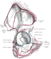
 Overview of Ilium as largest bone of the pelvis. | |
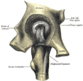
 Capsule of hip-joint (distended). Posterior aspect. (Ilium labeled at top.) | |
| Details | |
| Identifiers | |
| Latin | os ilii |
| MeSH | D007085 |
| TA98 | A02.5.01.101 |
| TA2 | 1317 |
| FMA | 16589 |
| Anatomical terms of bone | |
The ilium is the uppermost and largest bone of the pelvis, and appears in most vertebrates including mammals and birds, but not bony fish. All reptiles have an ilium except snakes, although some snake species have a tiny bone which is considered to be an ilium.[1]
The name comes from the Latin (ile, ilis), meaning "groin" or "flank."[2]
The ilium of the human is divisible into two parts, the body and the ala; the separation is indicated on the top surface by a curved line, the arcuate line, and on the external surface by the margin of the acetabulum.
Body (corpus oss. ilii)
The body enters into the formation of the acetabulum, of which it forms rather less than two-fifths.
Its external surface is partly articular, partly non-articular; the articular segment forms part of the lunate surface of the acetabulum, the non-articular portion contributes to the acetabular fossa.
The internal surface of the body is part of the wall of the lesser pelvis and gives origin to some fibers of the Obturator internus.
Below, it is continuous with the pelvic surfaces of the ischium and pubis, only a faint line indicating the place of union.
Ala (ala oss. ilii)
The wing of ilium (or ala) is the large expanded portion which bounds the greater pelvis laterally. It presents for examination two surfaces—an external and an internal—a crest, and two borders—an anterior and a posterior.
Biiliac width
In humans, biiliac width is an anatomical term referring to the widest measure of the pelvis between the outer edges of the upper iliac bones.
Biiliac width has the following common synonyms: pelvic bone width, biiliac breadth, intercristal breadth/width, bi-iliac breadth/width and biiliocristal breadth/width.
In the average adult female, it measures 28 cm (11 in).[citation needed] It is best measured by anthropometric calipers (an anthropometer designed for such measurement is called a pelvimeter). Attempting to measure biiliac width with a tape measure along a curved surface is inaccurate.
The biiliac width measure is helpful in obstetrics because a pelvis that is significantly too small or too large can have obstetrical complications. For example, a large baby and/or a small pelvis often lead to death unless a caesarean section is performed. [3]
It is also used by anthropologists to estimate body mass.[4]
See also
Additional images
-
Pelvic girdle
-
Male pelvis.
-
Right hip bone. Internal surface.
-
Right hip bone. External surface. (Body of ilium is the top of the blue circle in the center, and the wing of the ilium is the portion above that. Crest of ilium is labeled at top.)
-
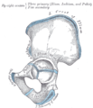
Plan of ossification of the hip bone.
-
Left hip-joint, opened by removing the floor of the acetabulum from within the pelvis.
-
Pelvis (French labels)
-
Pelvis
References
- ^
Jacobson, Elliott R. (2007). Infectious Diseases and Pathology of Reptiles. CRC Press. p. 7. ISBN 0849323215, 9780849323218. Retrieved 2009-01-09.
{{cite book}}: Check|isbn=value: invalid character (help) - ^ Taber, Clarence Wilbur; Venes, Donald (2005). Taber's cyclopedic medical dictionary. Philadelphia: F.A. Davis. ISBN 0-8036-1207-9.
{{cite book}}: CS1 maint: multiple names: authors list (link) - ^ "Encyclopedia of Medicine: Cesarean Section". eNotes.
- ^ Ruff C, Niskanenb M, Junnob J, Jamisonc P (2005). "Body mass prediction from stature and bi-iliac breadth in two high latitude populations, with application to earlier higher latitude humans" (PDF). Journal of Human Evolution. 48 (4): 381–392. doi:10.1016/j.jhevol.2004.11.009. PMID 15788184. Retrieved 2006-07-26.
{{cite journal}}: CS1 maint: multiple names: authors list (link)
External links
- Anatomy photo:44:st-0701 at the SUNY Downstate Medical Center
- pelvis at The Anatomy Lesson by Wesley Norman (Georgetown University)
![]() This article incorporates text in the public domain from page 236 of the 20th edition of Gray's Anatomy (1918)
This article incorporates text in the public domain from page 236 of the 20th edition of Gray's Anatomy (1918)


