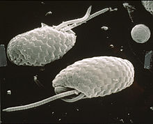
Mastigoneme


Mastigonemes are lateral "hairs" that attach to protistan flagella. Flimsy hairs attach to the flagella of euglenid flagellates, while stiff hairs occur in stramenopile and cryptophyte protists.[1] Stramenopile hairs are approximately 15 nm in diameter, and usually consist of flexible basal part that inserts into the cell membrane, a tubular shaft that itself terminates in smaller "hairs". They reverse the thrust caused when a flagellum beats. The consequence is that the cell is drawn into the water and particles of food are drawn to the surface of heterotrophic species.
Typology of flagella with hairs:[2][3][4][5][6]
- whiplash flagella (= smooth, acronematic flagella): without hairs but may have extensions , e.g., in Opisthokonta
- hairy flagella (= tinsel, flimmer, pleuronematic flagella): with hairs (= mastigonemes sensu lato), divided in:
- with fine hairs (= non tubular, or simple hairs): occurs in Euglenophyceae, Dinoflagellata, some Haptophyceae (Pavlovales)
- with stiff hairs (= tubular hairs, retronemes, mastigonemes sensu stricto), divided in:
- bipartite hairs: with two regions. Occurs in Cryptophyceae, Prasinophyceae, and some Heterokonta
- tripartite (= straminipilous) hairs: with three regions (a base, a tubular shaft, and one or more terminal hairs). Occurs in most Heterokonta/Stramenopiles
Observations of mastigonemes using light microscopy dates from the nineteenth century.[7][8][9][10][11] Considered artifacts by some, their existence would be confirmed with electron microscopy.[12][13]
References
[edit]- ^ Hoek, C. van den, Mann, D. G. and Jahns, H. M. (1995). Algae : An introduction to phycology, Cambridge University Press, UK.
- ^ Webster & Weber (2007).
- ^ South, G.R. & Whittick, A. (1987). Introduction to Phycology. Blackwell Scientific Publications, Oxford. p. 65, [1].
- ^ Barsanti, Laura; Gualtieri, Paolo (2006). Algae: anatomy, biochemistry, and biotechnology. Florida, USA: CRC Press. pp. 60-63, [2]
- ^ Dodge, J.D. (1973). The Fine Structure of Algal Cells. Academic Press, London. pp. 57-79, [3]
- ^ Lee, R. E. (2008). Phycology (4th ed.). Cambridge University Press. p. 7, [4].
- ^ Loeffler, F. (1889). Eine neue Methode zum Färbern der Mikroorganismen, im besonderen ihrer Wimperhaare und Geisseln. Zentralblatt für Bakteriologie und Parasitenkunde, 6, 209–224. [5].
- ^ Fischer, A. (1894). Über die Geißeln einiger Flagellaten. Jahrbuch für wissenchaftliche Botanik 26: 187-235.
- ^ Petersen, J. B. (1929). Beiträge zur Kenntnis der Flagellatengeißeln. Saertryk af Botanisk Tidsskrift. Bd. 40. 5. Heft.
- ^ Vlk, W. (1931). Uber die Struktur der Heterokontengeisseln. Botanisch Centralblatt 48: 214–220. [6].
- ^ Deflandre, G. (1934). Sur la structure des flagelles. Annales de Protistologie Vol. IV, pp. 31-54.
- ^ Pitelka, D. R. (1963). Electron-Microscopic Structure of Protozoa. Pergamon Press, Oxford. [7]
- ^ Bouck, G.B. 1971. The structure, origin, and comnposition of the tubular mastigonemes of the Ochromonas flagellum. J. Cell. Biol., 50: 362-384
