Posterior ethmoidal nerve
| Posterior ethmoidal nerve | |
|---|---|
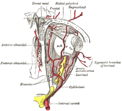
 The ophthalmic artery and its branches. (Nerve not pictured, but location is similar to artery.) | |
| Details | |
| From | nasociliary nerve |
| Innervates | sphenoidal sinus, ethmoidal sinus |
| Identifiers | |
| Latin | nervus ethmoidalis posterior |
| TA98 | A14.2.01.028 |
| TA2 | 6207 |
| FMA | 52714 |
| Anatomical terms of neuroanatomy | |
The posterior ethmoidal nerve is a nerve of the orbit around the eye. It is a branch of the nasociliary nerve from the ophthalmic nerve (CN V1). It supplies sensation to the sphenoid sinus, the ethmoid sinus, and part of the dura mater in the anterior cranial fossa.
Structure
The posterior ethmoidal nerve is a branch of the nasociliary nerve, itself a branch of the ophthalmic nerve (CN V1), itself a branch of the trigeminal nerve (CN V).[1] It passes through the posterior ethmoidal foramen, with the posterior ethmoidal artery.[2] It gives branches to the sphenoid sinus and the ethmoid sinus.[1] It also gives a branch to supply part of the dura mater in the anterior cranial fossa.[3][4]
Variation
The posterior ethmoidal nerve is absent in a significant proportion of people.[5] This may be around 30%.
Function
The posterior ethmoidal nerve supplies sensation to the sphenoid sinus and the ethmoid sinus.[1] It also supplies sensation to part of the dura mater in the anterior cranial fossa.[3][4]
Other animals
The posterior ethmoidal nerve is present in other animals, including horses.[6][7] Headshaking can sometimes be treated with analgesia or neurectomy of the posterior ethmoidal nerve.[7]
Reference
- ^ a b c Barral, Jean-Pierre; Croibier, Alain (2009). "15 - Ophthalmic nerve". Manual Therapy for the Cranial Nerves. Churchill Livingstone. pp. 115–128. doi:10.1016/B978-0-7020-3100-7.50018-5. ISBN 978-0-7020-3100-7.
- ^ Semmer, A. E.; McLoon, L. K.; Lee, M. S. (2010). "Orbital Vascular Anatomy". Encyclopedia of the Eye. Academic Press. pp. 241–251. doi:10.1016/B978-0-12-374203-2.00284-0. ISBN 978-0-12-374203-2.
- ^ a b Shimizu, Toshihiko; Suzuki, Norihiro (2010). "3 - Biological sciences related to headache". Handbook of Clinical Neurology. Vol. 97. Elsevier. pp. 35–45. doi:10.1016/S0072-9752(10)97003-6. ISBN 978-0-444-52139-2. ISSN 0072-9752.
- ^ a b Seker, Askin; Martins, Carolina; Rhoton Jr., Albert L. (2010). "2 - Meningeal Anatomy". Meningiomas. Saunders. pp. 11–51. doi:10.1016/B978-1-4160-5654-6.00002-7. ISBN 978-1-4160-5654-6.
- ^ Rea, Paul (2016). "2 - Head". Essential Clinically Applied Anatomy of the Peripheral Nervous System in the Head and Neck. Academic Press. pp. 21–130. doi:10.1016/B978-0-12-803633-4.00002-8. ISBN 978-0-12-803633-4.
- ^ "11 - Disorders of the nervous system". Knottenbelt and Pascoe's Color Atlas of Diseases and Disorders of the Horse (2nd ed.). Saunders. 2014. pp. 400–442. doi:10.1016/B978-0-7234-3660-7.00011-0. ISBN 978-0-7234-3660-7.
- ^ a b Carr, Elizabeth A.; Maher, Omar (2014). Equine Sports Medicine and Surgery (2nd ed.). Saunders. pp. 503–526. doi:10.1016/B978-0-7020-4771-8.00024-7. ISBN 978-0-7020-4771-8.
External links
- MedEd at Loyola GrossAnatomy/h_n/cn/cn1/cnb1.htm
- cranialnerves at The Anatomy Lesson by Wesley Norman (Georgetown University) (V)
- http://www.dartmouth.edu/~humananatomy/figures/chapter_45/45-6.HTM
