
Subclavian vein: Difference between revisions
Fixing spelling Tag: Mobile edit |
|||
| Line 6: | Line 6: | ||
Image = Gray1174.png | |
Image = Gray1174.png | |
||
Caption = The [[thyroid gland]] and its relations. (Right subclavian vein visible at bottom left, left subclavian vein visible at bottom right.) | |
Caption = The [[thyroid gland]] and its relations. (Right subclavian vein visible at bottom left, left subclavian vein visible at bottom right.) | |
||
Image2 = |
Image2 = Gray40.png | |
||
Caption2 = Diagram showing completion of development of the [[parietal veins]]. (Subclavian vein labeled at right, third from top.) | |
Caption2 = Diagram showing completion of development of the [[parietal veins]]. (Subclavian vein labeled at right, third from top.) | |
||
DrainsFrom = | |
DrainsFrom = | |
||
| Line 35: | Line 35: | ||
==Additional images== |
==Additional images== |
||
<gallery> |
<gallery> |
||
Image: |
Image:Gray12.png|Peculiar ribs. |
||
Image:Venenwinkel.png|The venæ cavæ and azygos veins, with their tributaries. |
Image:Venenwinkel.png|The venæ cavæ and azygos veins, with their tributaries. |
||
Image: |
Image:Gray590.png|The thoracic and right lymphaticducts. |
||
Image: |
Image:Gray178.png|The thymus of a full-term fetus, exposed in situ. |
||
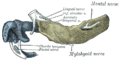
File:Slide9a.JPG|Subclavian vein |
File:Slide9a.JPG|Subclavian vein |
||
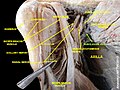
File:Slide7d.JPG|Subclavian vein - right view |
File:Slide7d.JPG|Subclavian vein - right view |
||
Revision as of 15:53, 19 March 2013
| Subclavian vein | |
|---|---|
 The thyroid gland and its relations. (Right subclavian vein visible at bottom left, left subclavian vein visible at bottom right.) | |
 Diagram showing completion of development of the parietal veins. (Subclavian vein labeled at right, third from top.) | |
| Details | |
| Source | axillary vein, external jugular vein |
| Drains to | brachiocephalic vein |
| Artery | subclavian artery |
| Identifiers | |
| Latin | vena subclavia |
| MeSH | D013350 |
| TA98 | A12.3.08.002 |
| TA2 | 4953 |
| FMA | 4725 |
| Anatomical terminology | |
The subclavian veins are two large veins, one on either side of the body. Their diameter is approximately that of the smallest finger.
Path
Each subclavian vein is a continuation of the axillary vein and runs from the outer border of the first rib to the medial border of anterior scalene muscle. From here it joins with the internal jugular vein to form the brachiocephalic vein (also known as "innominate vein"). The angle of union is termed the venous angle.
The subclavian vein follows the subclavian artery and is separated from the subclavian artery by the insertion of anterior scalene. Thus, the subclavian vein lies anterior to the anterior scalene while the subclavian artery lies posterior to the anterior scalene (and anterior to the middle scalene).
Lymph
The thoracic duct drains into the left subclavian vein, near its junction with the left internal jugular vein. It carries lymph (water and solutes) from the lymphatic system, as well as chylomicrons or chyle, formed in the intestines from dietary fat and lipids.
The right lymphatic duct drains its lymph into the junction of the right internal jugular vein, and the right subclavian vein.
Etymology
Sub (below), and clavian (pertaining to the clavicle).
See also
Additional images
-
 Peculiar ribs.
 -
 The venæ cavæ and azygos veins, with their tributaries.
 -
 The thoracic and right lymphaticducts.
 -
 The thymus of a full-term fetus, exposed in situ.
 -
 Subclavian vein
 -
 Subclavian vein - right view
 -
 Subclavian vein
 -
 Subclavian vein


External links
This cardiovascular system article is a stub. You can help Wikipedia by expanding it. |