Cerebral infarction
| Cerebral infarct | |
|---|---|
 | |
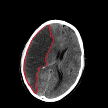
| CT scan slice of the brain showing a right-hemispheric cerebral infarct (left side of image). | |
| Specialty | Neurology |
Cerebral infarction, also known as an ischemic stroke, is the pathologic process that results in an area of necrotic tissue in the brain (cerebral infarct).[1] In mid to high income countries, a stroke is the main reason for disability among people and the 2nd cause of death.[2] It is caused by disrupted blood supply (ischemia) and restricted oxygen supply (hypoxia). This is most commonly due to a thrombotic occlusion, or an embolic occlusion of major vessels which leads to a cerebral infarct .[3][4] In response to ischemia, the brain degenerates by the process of liquefactive necrosis.[1]
Classification
[edit]There are various classification systems[5] for cerebral infarcts, some of which are described below.
- The Oxford Community Stroke Project classification (OCSP, also known as the Bamford or Oxford classification) relies primarily on the initial symptoms. Based on the extent of the symptoms, the stroke episode is classified as total anterior circulation infarct (TACI), partial anterior circulation infarct (PACI), lacunar infarct (LACI) or posterior circulation infarct (POCI). These four entities predict the extent of the stroke, the area of the brain affected, the underlying cause, and the prognosis.[6][7]
- The TOAST (Trial of Org 10172 in Acute Stroke Treatment) classification is based on clinical symptoms as well as results of further investigations; on this basis, a stroke is classified as being due to (1) thrombosis or embolism due to atherosclerosis of a large artery, (2) embolism of cardiac origin, (3) occlusion of a small blood vessel, (4) other determined cause, (5) undetermined cause (two possible causes, no cause identified, or incomplete investigation).[8]
Stroke recognition
[edit]There are many tests that can be done to prescreen a patient who may be showing stroke-like symptoms. No one test is better than the other and they all have room for improvement. One of these tests that is used by prehospital personnel is the Cincinnati Pre Hospital Stroke scale (CPSS). This test looks for facial droop, arm drift, and a change in the person's speech pattern. Another test that can be used and is a modification to the CPSS is the Face Arm Speech Test (FAST). This checks for facial weakness, arm weakness, and speech disturbances. The ROSIER (Recognition of Stroke in The ER), is a test used by an ER physician. Each variable is rated from a -2 to a +5. Loss of consciousness (-1), convulsive fit (-1), facial weakness (+1), arm weakness (+1), leg weakness (+1), abnormal speech patterns (+1), and visual defect (+1).[9] In recent years, a study has been done to show how AI can aid in diagnosis of cerebral infarct and improve patient outcomes in areas that may not have stroke trained physicians.[10]
Symptoms
[edit]


Ischemic strokes usually present as a problem with nerve, spinal cord, or brain function. Depending on where the stroke is located in the brain, symptoms may start within minutes, or they make take hours to present themselves. Most strokes occur without warning. Some common symptoms include one sided weakness, facial paralysis or numbness, vision problems, trouble speaking, problems with walking and keeping balanced. A person can show one or more of these symptoms during a stroke. If person has a decrease in consciousness, they may be suffering from a stroke in more than one part of the brain or in the brain stem.[12]
Symptoms of cerebral infarction can help determine which parts of the brain are affected. If the infarct is located in the primary motor cortex, contralateral hemiparesis is said to occur. With brainstem localization, brainstem syndromes are typical: Wallenberg's syndrome, Weber's syndrome, Millard–Gubler syndrome, Benedikt syndrome or others.
Risk factors
[edit]Major risk factors for cerebral infarction are generally the same as for atherosclerosis. These include high blood pressure, diabetes mellitus, tobacco smoking, obesity, and dyslipidemia.[13] There are also risks that a person can't control. These include a person's age, family history of strokes, being African American, and being born a male. A person's risk of a stroke doubles each decade after the age of 55.[14] The American Heart Association/American Stroke Association (AHA/ASA) recommends controlling these risk factors in order to prevent stroke.[15] The AHA/ASA guidelines also provide information on how to prevent stroke if someone has more specific concerns, such as sickle-cell disease or pregnancy. It is also possible to calculate the risk of stroke in the next decade based on information gathered through the Framingham Heart Study.[16]
Pathophysiology
[edit]Cerebral infarction is caused by a disruption to blood supply that is severe enough and long enough in duration to result in tissue death. The disruption to blood supply can come from many causes, including:
- Thrombosis (obstruction of a blood vessel by a blood clot forming locally)
- Embolism (obstruction due to an embolus from elsewhere in the body),[17]
- Systemic hypoperfusion (general decrease in blood supply, e.g., in shock)[18]
- Cerebral venous sinus thrombosis.[19]
- Unusual causes such as gas embolism from rapid ascents in scuba diving.[20]
Even in cases where there is a complete blockage to blood flow of a major blood vessel supplying the brain, there is typically some blood flow to the downstream tissue through collateral blood vessels, and the tissue can typically survive for some length of time that is dependent upon the level of remaining blood flow.[21] If blood flow is reduced enough, oxygen delivery can decrease enough to cause the tissue to undergo the ischemic cascade. The ischemic cascade leads to energy failure that prevents neurons from sufficiently moving ions through active transport which leads the neurons to first cease firing, then depolarize leading to ion imbalances that cause fluid inflows and cellular edema, then undergo a complex chain of events that can lead to cell death through one or more pathways.[22][23][24]
Diagnosis
[edit]Computed tomography (CT) and MRI scanning will show damaged area in the brain. A CT scan will rule out a hemorrhagic stroke, is cheaper for the patient, and can be found in almost all hospitals unlike an MRI machine.[25][26] Once the Doctor rules out a hemorrhagic stroke, rTPA can be given.[25] An MRI can help to diagnose an acute cerebral infarct as quickly as 6 hours from start of symptoms,[25] It can also help time when the stroke happened.[27] The biggest problem with an MRI is it can't be done on a patient with certain metallic implants or if the patient is claustrophobic.[28] A head and neck CT angiogram can be performed within 6 hours of onset of symptoms to see where the occlusion may be located which can help in determining the cause of the stroke.[29] In people who die from a stroke an autopsy can reveal additional diseases or conditions beyond the stroke itself, as well as uncover uncommon causes of a stroke.[30]
Treatment
[edit]In the last decade, similar to myocardial infarction treatment, thrombolytic drugs were introduced in the therapy of cerebral infarction. The use of intravenous rtPA therapy can be advocated in patients who arrive to stroke unit and can be fully evaluated within 3 hours of the onset. The quicker rTPA is started, the better the outcome for the patient.[29]
If cerebral infarction is caused by a thrombus occluding blood flow to an artery supplying the brain, definitive therapy is aimed at removing the blockage by breaking the clot down (thrombolysis), or by removing it mechanically (thrombectomy). The more rapidly blood flow is restored to the brain, the fewer brain cells die.[31] In increasing numbers of primary stroke centers, pharmacologic thrombolysis with the drug tissue plasminogen activator (tPA), is used to dissolve the clot and unblock the artery. Giving rTPA lessens the chance of disability after 3 months by 30%.[29] Another intervention for acute cerebral ischemia is removal of the offending thrombus directly. This is accomplished by inserting a catheter into the femoral artery, directing it into the cerebral circulation, and deploying a corkscrew-like device to ensnare the clot, which is then withdrawn from the body. Mechanical embolectomy devices have been demonstrated effective at restoring blood flow in patients who were unable to receive thrombolytic drugs or for whom the drugs were ineffective,[32][33][34][35] though no differences have been found between newer and older versions of the devices.[36] The devices have only been tested on patients treated with mechanical clot embolectomy within eight hours of the onset of symptoms.
Angioplasty and stenting have begun to be looked at as possible viable options in treatment of acute cerebral ischaemia. In a systematic review of six uncontrolled, single-center trials, involving a total of 300 patients, of intra-cranial stenting in symptomatic intracranial arterial stenosis, the rate of technical success (reduction to stenosis of <50%) ranged from 90 to 98%, and the rate of major peri-procedural complications ranged from 4-10%. The rates of restenosis and/or stroke following the treatment were also favorable.[37] This data suggests that a large, randomized controlled trial is needed to more completely evaluate the possible therapeutic advantage of this treatment.
If studies show carotid stenosis, and the patient has residual function in the affected side, carotid endarterectomy (surgical removal of the stenosis) may decrease the risk of recurrence if performed rapidly after cerebral infarction. Carotid endarterectomy is also indicated to decrease the risk of cerebral infarction for symptomatic carotid stenosis (>70 to 80% reduction in diameter).[38]
In tissue losses that are not immediately fatal, the best course of action is to make every effort to restore impairments through physical therapy, cognitive therapy, occupational therapy, speech therapy and exercise.
Permissive hypertension - allowing for higher than normal blood pressures in the acute phase of cerebral infarction - can be used to encourage perfusion to the penumbra.[39]
References
[edit]- ^ a b Kumar V, Abbas AK, Aster JC, Perkins JA, Robbins SL, Cotran RS (2015). Robbins and Cotran pathologic basis of disease. Student consult (Ninth ed.). Philadelphia, Pa: Elsevier; Saunders. ISBN 978-1-4557-2613-4.
- ^ Murphy SJ, Werring DJ (September 2020). "Stroke: causes and clinical features". Medicine. 48 (9): 561–566. doi:10.1016/j.mpmed.2020.06.002. PMC 7409792. PMID 32837228.
- ^ Douglas VC, Aminoff MJ (2023). "24-09: Stroke". In Papadakis MA, McPhee SJ, Rabow MW, McQuaid KR (eds.). Current Medical Diagnosis & Treatment 2023. New York, NY: McGraw-Hill Education. Retrieved 2024-03-21.
- ^ Shin TH, Lee DY, Basith S, Manavalan B, Paik MJ, Rybinnik I, et al. (July 2020). "Metabolome Changes in Cerebral Ischemia". Cells. 9 (7): 1630. doi:10.3390/cells9071630. PMC 7407387. PMID 32645907.
- ^ Amarenco P, Bogousslavsky J, Caplan LR, Donnan GA, Hennerici MG (2009). "Classification of stroke subtypes". Cerebrovascular Diseases. 27 (5): 493–501. doi:10.1159/000210432. PMID 19342825.
- ^ Bamford J, Sandercock P, Dennis M, Burn J, Warlow C (June 1991). "Classification and natural history of clinically identifiable subtypes of cerebral infarction". Lancet. 337 (8756): 1521–1526. doi:10.1016/0140-6736(91)93206-O. PMID 1675378. S2CID 21784682. Later publications distinguish between "syndrome" and "infarct", based on evidence from imaging. "Syndrome" may be replaced by "hemorrhage" if imaging demonstrates a bleed. See Internet Stroke Center. "Oxford Stroke Scale". Retrieved 2008-11-14.
- ^ Bamford JM (2000). "The role of the clinical examination in the subclassification of stroke". Cerebrovascular Diseases. 10 (4): 2–4. doi:10.1159/000047582. PMID 11070389. S2CID 29493084.
- ^ Adams HP, Bendixen BH, Kappelle LJ, Biller J, Love BB, Gordon DL, et al. (January 1993). "Classification of subtype of acute ischemic stroke. Definitions for use in a multicenter clinical trial. TOAST. Trial of Org 10172 in Acute Stroke Treatment". Stroke. 24 (1): 35–41. doi:10.1161/01.STR.24.1.35. PMID 7678184.
- ^ Chaudhary D, Diaz J, Lu Y, Li J, Abedi V, Zand R (November 2022). "An updated review and meta-analysis of screening tools for stroke in the emergency room and prehospital setting". Journal of the Neurological Sciences. 442: 120423. doi:10.1016/j.jns.2022.120423. PMID 36201961.
- ^ Miyamoto N, Ueno Y, Yamashiro K, Hira K, Kijima C, Kitora N, et al. (2023-12-14). "Stroke classification and treatment support system artificial intelligence for usefulness of stroke diagnosis". Frontiers in Neurology. 14: 1295642. doi:10.3389/fneur.2023.1295642. PMC 10753815. PMID 38156087.
- ^ Kentar M, Mann M, Sahm F, Olivares-Rivera A, Sanchez-Porras R, Zerelles R, et al. (March 2020). "Detection of spreading depolarizations in a middle cerebral artery occlusion model in swine". Acta Neurochirurgica. 162 (3): 581–592. doi:10.1007/s00701-019-04132-8. PMID 31940093. S2CID 210196036.
- ^ Lyerly M (2020). "Chapter 24: Stroke & Neurovascular Disorders". In Amthor F, Theibert WA, Standaert DG, Roberson E (eds.). Essentials of Modern Neuroscience. New York, NY: McGraw Hill LLC. p. 7. ISBN 978-1-259-86104-8.
- ^ Hankey GJ (August 2006). "Potential new risk factors for ischemic stroke: what is their potential?". Stroke. 37 (8): 2181–2188. doi:10.1161/01.STR.0000229883.72010.e4. PMID 16809576.
- ^ Powers WJ, Rabinstein AA, Ackerson T, Adeoye OM, Bambakidis NC, Becker K, et al. (March 2018). "2018 Guidelines for the Early Management of Patients With Acute Ischemic Stroke: A Guideline for Healthcare Professionals From the American Heart Association/American Stroke Association". Stroke. 49 (3): e46–e110. doi:10.1161/STR.0000000000000158. PMID 29367334.
- ^ Furie KL, Kasner SE, Adams RJ, Albers GW, Bush RL, Fagan SC, et al. (January 2011). "Guidelines for the prevention of stroke in patients with stroke or transient ischemic attack: a guideline for healthcare professionals from the american heart association/american stroke association". Stroke. 42 (1): 227–276. doi:10.1161/STR.0b013e3181f7d043. PMID 20966421.
- ^ D'Agostino RB, Wolf PA, Belanger AJ, Kannel WB (January 1994). "Stroke risk profile: adjustment for antihypertensive medication. The Framingham Study". Stroke. 25 (1): 40–43. doi:10.1161/01.str.25.1.40. PMID 8266381.
- ^ Donnan GA, Fisher M, Macleod M, Davis SM (May 2008). "Stroke". Lancet. 371 (9624): 1612–1623. doi:10.1016/S0140-6736(08)60694-7. PMID 18468545. S2CID 208787942.(subscription required)
- ^ Shuaib A, Hachinski VC (September 1991). "Mechanisms and management of stroke in the elderly". CMAJ. 145 (5): 433–443. PMC 1335826. PMID 1878825.
- ^ Stam J (April 2005). "Thrombosis of the cerebral veins and sinuses". The New England Journal of Medicine. 352 (17): 1791–1798. doi:10.1056/NEJMra042354. PMID 15858188. S2CID 42126852.
- ^ Levett DZ, Millar IL (November 2008). "Bubble trouble: a review of diving physiology and disease". Postgraduate Medical Journal. 84 (997): 571–578. doi:10.1136/pgmj.2008.068320. PMID 19103814. S2CID 1165852.
- ^ Vagal A, Aviv R, Sucharew H, Reddy M, Hou Q, Michel P, et al. (September 2018). "Collateral Clock Is More Important Than Time Clock for Tissue Fate". Stroke. 49 (9): 2102–2107. doi:10.1161/STROKEAHA.118.021484. PMC 6206882. PMID 30354992.
- ^ Krnjević K (September 1999). "Early effects of hypoxia on brain cell function". Croatian Medical Journal. 40 (3): 375–380. PMID 10411965.
- ^ Hartings JA, Shuttleworth CW, Kirov SA, Ayata C, Hinzman JM, Foreman B, et al. (May 2017). "The continuum of spreading depolarizations in acute cortical lesion development: Examining Leão's legacy". Journal of Cerebral Blood Flow and Metabolism. 37 (5): 1571–1594. doi:10.1177/0271678X16654495. PMC 5435288. PMID 27328690.
- ^ Kalogeris T, Baines CP, Krenz M, Korthuis RJ (December 2016). "Ischemia/Reperfusion". Comprehensive Physiology. 7 (1): 113–170. doi:10.1002/cphy.c160006. ISBN 978-0-470-65071-4. PMC 5648017. PMID 28135002.
- ^ a b c Lin MP, Liebeskind DS (October 2016). "Imaging of Ischemic Stroke". Continuum. 22 (5, Neuroimaging): 1399–1423. doi:10.1212/CON.0000000000000376. PMC 5898964. PMID 27740982.
- ^ Hakimi R, Garg A (October 2016). "Imaging of Hemorrhagic Stroke". Continuum. 22 (5, Neuroimaging): 1424–1450. doi:10.1212/CON.0000000000000377. PMID 27740983.
- ^ White P, Nanapragasam A (April 2018). "What is new in stroke imaging and intervention?". Clinical Medicine. 18 (Suppl 2): s13–s16. doi:10.7861/clinmedicine.18-2-s13. PMC 6334026. PMID 29700087.
- ^ Zhang XH, Liang HM (July 2019). "Systematic review with network meta-analysis: Diagnostic values of ultrasonography, computed tomography, and magnetic resonance imaging in patients with ischemic stroke". Medicine. 98 (30): e16360. doi:10.1097/MD.0000000000016360. PMC 6709059. PMID 31348236.
- ^ a b c Papadakis M, McPhee SJ (2024). "Stroke, Cerebral Infarction". Quick Medical Diagnosis & Treatment 2024. New York, NY: McGraw-Hill Education. Retrieved 2024-03-25.
- ^ Hudák L, Nagy AC, Molnár S, Méhes G, Nagy KE, Oláh L, et al. (June 2022). "Discrepancies between clinical and autopsy findings in patients who had an acute stroke". Stroke and Vascular Neurology. 7 (3): 215–221. doi:10.1136/svn-2021-001030. PMC 9240455. PMID 35101949.
- ^ Saver JL (January 2006). "Time is brain--quantified". Stroke. 37 (1): 263–266. doi:10.1161/01.STR.0000196957.55928.ab. PMID 16339467.
- ^ Flint AC, Duckwiler GR, Budzik RF, Liebeskind DS, Smith WS (April 2007). "Mechanical thrombectomy of intracranial internal carotid occlusion: pooled results of the MERCI and Multi MERCI Part I trials". Stroke. 38 (4): 1274–1280. doi:10.1161/01.STR.0000260187.33864.a7. PMID 17332445.
- ^ Smith WS, Sung G, Starkman S, Saver JL, Kidwell CS, Gobin YP, et al. (July 2005). "Safety and efficacy of mechanical embolectomy in acute ischemic stroke: results of the MERCI trial". Stroke. 36 (7): 1432–1438. doi:10.1161/01.STR.0000171066.25248.1d. PMID 15961709.
- ^ Lutsep HL, Rymer MM, Nesbit GM (2008). "Vertebrobasilar revascularization rates and outcomes in the MERCI and multi-MERCI trials". Journal of Stroke and Cerebrovascular Diseases. 17 (2): 55–57. doi:10.1016/j.jstrokecerebrovasdis.2007.11.003. PMID 18346645.
- ^ Smith WS (June 1, 2006). "Safety of mechanical thrombectomy and intravenous tissue plasminogen activator in acute ischemic stroke. Results of the multi Mechanical Embolus Removal in Cerebral Ischemia (MERCI) trial, part I". AJNR. American Journal of Neuroradiology. 27 (6): 1177–1182. PMC 8133930. PMID 16775259.
- ^ Smith WS, Sung G, Saver J, Budzik R, Duckwiler G, Liebeskind DS, et al. (April 2008). "Mechanical thrombectomy for acute ischemic stroke: final results of the Multi MERCI trial". Stroke. 39 (4): 1205–1212. doi:10.1161/STROKEAHA.107.497115. PMID 18309168.
- ^ Derdeyn CP, Chimowitz MI (August 2007). "Angioplasty and stenting for atherosclerotic intracranial stenosis: rationale for a randomized clinical trial". Neuroimaging Clinics of North America. 17 (3): 355–63, viii–ix. doi:10.1016/j.nic.2007.05.001. PMC 2040119. PMID 17826637.
- ^ Ropper AH, Brown RH, eds. (2005). Adams and Victor's Principles of Neurology (8th ed.). McGraw Hill Professional. p. 698. ISBN 0-07-141620-X.
- ^ Grotta JC, Albers G, Broderick JP, Kasner SE, Lo EH, Mendelow AD, eds. (24 August 2015). Stroke: pathophysiology, diagnosis, and management (Sixth ed.). Elsevier. ISBN 978-0-323-29544-4. OCLC 919749088.
