Dorsum sellae
Appearance
| Dorsum sellae | |
|---|---|
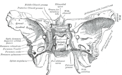
 Sphenoid bone. Upper surface. (Dorsum sellae is labeled in the white portion in the center.) | |
 Base of the skull. Upper surface. (Dorsum sellae is not labeled, but sphenoid bone is in yellow, and Dorsum sellae would horizontally be at the exact center, and vertically at the level of the posterior clinoid process.) | |
| Details | |
| Identifiers | |
| Latin | dorsum sellae, dorsum sella turcica |
| TA98 | A02.1.05.010 |
| TA2 | 594 |
| FMA | 54718 |
| Anatomical terms of bone | |
The dorsum sellae is part of the sphenoid bone in the skull. Together with the basilar part of the occipital bone it forms the clivus.
In the sphenoid bone, the anterior boundary of the sella turcica is completed by two small eminences, one on either side, called the middle clinoid processes, while the posterior boundary is formed by a square-shaped plate of bone, the dorsum sellae, ending at its superior angles in two tubercles, the posterior clinoid processes, the size and form of which vary considerably in different individuals.
Additional images
[edit]-
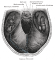
Tentorium cerebelli from above.
-
Dorsum sellae
References
[edit]![]() This article incorporates text in the public domain from page 147 of the 20th edition of Gray's Anatomy (1918)
This article incorporates text in the public domain from page 147 of the 20th edition of Gray's Anatomy (1918)
External links
[edit]- "Anatomy diagram: 34257.000-2". Roche Lexicon - illustrated navigator. Elsevier. Archived from the original on 2013-06-22.
- Anatomy photo:22:os-0912 at the SUNY Downstate Medical Center


