
Pulmonary vein
| Pulmonary vein | |
|---|---|
 Diagram of the alveoli with both cross-section and external view. | |
| Details | |
| Precursor | truncus arteriosus |
| Drains from | lungs |
| Drains to | left atrium |
| Artery | pulmonary artery |
| Identifiers | |
| Latin | venae pulmonales |
| MeSH | D011667 |
| TA98 | A12.3.02.001 |
| TA2 | 4107 |
| FMA | 66643 |
| Anatomical terminology | |
The pulmonary veins are large blood vessels that carry oxygenated blood from the lungs to the left atrium of the heart. In humans there are four pulmonary veins, two from each lung. They carry oxygenated blood, which is unusual since almost all other veins carry deoxygenated blood.
Path
Occasionally the three veins on the right side remain separate, and not infrequently the two left pulmonary veins end by a common opening into the left atrium. Therefore, the number of pulmonary veins opening into the left atrium can vary between three and five in the healthy population.
At the root of the lung, the superior pulmonary vein lies in front of and a little below the pulmonary artery; the inferior is situated at the lowest part of the hilus of the lung and on a plane posterior to the upper vein. Behind the pulmonary artery is the bronchus.
Within the pericardium, their anterior surfaces are invested by the serous layer of this membrane.
The right pulmonary veins pass behind the right atrium and superior vena cava; the left in front of the descending thoracic aorta.
Additional images
-
 Bronchial anatomy
 -
 Bronchi, bronchial tree, and lungs
 -
 Pulmonary circuit
 -
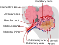 Alveolus diagram
 -
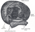 Heart seen from above.
 -
 Transverse section of thorax, showing relations of pulmonary artery.
 -
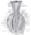
 Pulmonary vessels, seen in a dorsal view of the heart and lungs.
 -
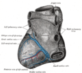 Base and diaphragmatic surface of heart.
 -
 The position and relation of the esophagus in the cervical region and in the posterior mediastinum. Seen from behind.
 -
 Left atrium


External links
- Anatomy figure: 19:05-08 at Human Anatomy Online, SUNY Downstate Medical Center
- Illustration at infomat.net
![]() This article incorporates text in the public domain from page 642 of the 20th edition of Gray's Anatomy (1918)
This article incorporates text in the public domain from page 642 of the 20th edition of Gray's Anatomy (1918)