Shigella
| Shigella | |
|---|---|

| |
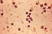
| Photomicrograph of Shigella sp. in a stool specimen | |
| Scientific classification | |
| Kingdom: | |
| Phylum: | |
| Class: | |
| Order: | |
| Family: | |
| Genus: | Shigella Castellani & Chalmers 1919
|
| Species | |
- This article is about the genus. For the disease, see shigellosis
Shigella is a genus of Gram-negative, nonspore forming, non-motile, rod-shaped bacteria closely related to Escherichia coli and Salmonella. The causative agent of human shigellosis, Shigella causes disease in primates, but not in other mammals.[1][page needed] It is only naturally found in humans and apes.[2] During infection, it typically causes dysentery.[3] The genus is named after Kiyoshi Shiga, who first discovered it in 1898.
Phylogenetic studies indicate that Shigella is more appropriately treated as subgenus of Escherichia, and that certain strains generally considered E. coli – such as E. coli O157:H7 – are better placed in Shigella (see Escherichia coli#Diversity for details).
Classification
Shigella species are classified by four serogroups:
- Serogroup A: S. dysenteriae (12 serotypes)
- Serogroup B: S. flexneri (6 serotypes)
- Serogroup C: S. boydii (18 serotypes)
- Serogroup D: S. sonnei (1 serotype)
Groups A–C are physiologically similar; S. sonnei (group D) can be differentiated on the basis of biochemical metabolism assays.[4] Three Shigella groups are the major disease-causing species: S. flexneri is the most frequently isolated species worldwide, and accounts for 60% of cases in the developing world; S. sonnei causes 77% of cases in the developed world, compared to only 15% of cases in the developing world; and S. dysenteriae is usually the cause of epidemics of dysentery, particularly in confined populations such as refugee camps.[5]
Pathogenesis
Shigella infection is typically via ingestion (fecal–oral contamination); depending on age and condition of the host, less than 100 bacterial cells can be enough to cause an infection.[6] Shigella causes dysentery that results in the destruction of the epithelial cells of the intestinal mucosa in the cecum and rectum. Some strains produce enterotoxin and shiga toxin, similar to the verotoxin of E. coli O157:H7[4] and other verotoxin-producing Escherichia coli. Both shiga toxin and verotoxin are associated with causing hemolytic uremic syndrome. As noted above, these supposed E. coli strains are at least in part actually more closely related to Shigella than to the "typical" E. coli.
Shigella invade the host through the M-cells in the gut epithelia of the large intestine, as they cannot enter directly through the epithelial cells. Using a Type III secretion system acting as a biological syringe, the bacterium injects IpaD protein into cells, triggering bacterial invasion and the subsequent lysis of vacuolar membranes using IpaB and IpaC proteins. It uses a mechanism for its motility by which its IcsA protein triggers actin polymerization in the host cell (via N-WASP recruitment of Arp2/3 complexes) in a "rocket" propulsion fashion for cell-to-cell spread. The most common symptoms are diarrhea, fever, nausea, vomiting, stomach cramps and flatulence. The stool may contain blood, mucus, or pus. In rare cases, young children may have seizures. Symptoms can take as long as a week to show up, but most often begin two to four days after ingestion. Symptoms usually last for several days, but can last for weeks. Shigella is implicated as one of the pathogenic causes of reactive arthritis worldwide.[7]
Each of the Shigella genomes includes a virulence plasmid that encodes conserved primary virulence determinants. The Shigella chromosomes share most of their genes with those of E. coli K12 strain MG1655.[8]
Diagnosis
Shigella species are negative for motility and are not lactose fermenters. (However, S. sonnei can ferment lactose).[9] They typically do not produce gas from carbohydrates (with the exception of certain strains of S. flexneri) and tend to be overall biochemically inert. Shigella should also be urea hydrolysis negative . When inoculated to a triple sugar iron (TSI) slant, they react as follows: K/A, gas -, H2S -. Indole reactions are mixed, positive and negative, with the exception of S. sonnei, which is always indole negative. Growth on Hektoen enteric agar will produce bluish-green colonies for Shigella and bluish-green colonies with black centers for Salmonella.
Prevention and Treatment
Hand washing before handling food and thoroughly cooking all food before eating decreases the risk of getting Shigella.[10]
Severe dysentery can be treated with ampicillin, TMP-SMX, or fluoroquinolones, such as ciprofloxacin, and of course rehydration. Medical treatment should only be used in severe cases. Antibiotics are usually avoided in mild cases because some Shigella are resistant to antibiotics, and their use may make the germ even more resistant. Antidiarrheal agents may worsen the sickness, and should be avoided.[11] For Shigella-associated diarrhea, antibiotics shorten the length of infection.[12]
Shigella is one of the leading bacterial causes of diarrhea worldwide. Insufficient data exist, but conservative estimates suggest that Shigella causes approximately 90 million cases of severe dysentery with at least 100,000 of these resulting in death each year, mostly among children in the developing world.[5]
Currently, no licenced vaccine targeting Shigella exists. Shigella has been a longstanding World Health Organization target for vaccine development, and sharp declines in age-specific diarrhea/dysentery attack rates for this pathogen indicate that natural immunity does develop following exposure; thus, vaccination to prevent the disease should be feasible. Several vaccine candidates for Shigella are in various stages of development.[5]
References
- ^ Ryan, Kenneth James; Ray, C. George, ed. (2004). Sherris medical microbiology: an introduction to infectious diseases (4 ed.). McGraw-Hill Professional Med/Tech. ISBN 978-0-8385-8529-0.
{{cite book}}: CS1 maint: multiple names: editors list (link) - ^ Attention: This template ({{cite doi}}) is deprecated. To cite the publication identified by doi:10.1007/s10669-006-8666-3, please use {{cite journal}} (if it was published in a bona fide academic journal, otherwise {{cite report}} with
|doi=10.1007/s10669-006-8666-3instead. - ^ Mims, Playfair, Roitt, Wakelin, Williams (1993). Medical Microbiology (1st ed.). Mosby. p. A.24. ISBN 0-397-44631-4.
{{cite book}}: CS1 maint: multiple names: authors list (link) - ^ a b Hale, Thomas L.; Keusch, Gerald T. (1996). "Shigella: Structure, Classification, and Antigenic Types". In Baron, Samuel (ed.). Medical microbiology (4 ed.). Galveston, Texas: University of Texas Medical Branch. ISBN 978-0-9631172-1-2. Retrieved February 11, 2012.
- ^ a b c "Diarrhoeal Diseases: Shigellosis". Initiative for Vaccine Research (IVR). World Health Organization. Retrieved May 11, 2012.
- ^ Levinson, Warren E (2006). Review of Medical Microbiology and Immunology (9 ed.). McGraw-Hill Medical Publishing Division. p. 30. ISBN 978-0-07-146031-6. Retrieved February 27, 2012.
- ^ Hill Gaston, J.S; Lillicrap, Mark S (April 2003). "Arthritis associated with enteric infection". Best Practice & Research. Clinical Rheumatology. 17 (2): 219–239. doi:10.1016/S1521-6942(02)00104-3. PMID 12787523.
- ^ Yang, Fan (2005). "Genome dynamics and diversity of Shigella species, the etiologic agents of bacillary dysentery" (PDF). Nucleic Acids Research. 33 (19): 6445–6458. doi:10.1093/nar/gki954. PMC 1278947. PMID 16275786. Retrieved February 11, 2012.
- ^ Ito H, Kido N, Arakawa Y, Ohta M, Sugiyama T, Kato N (1991). "Possible mechanisms underlying the slow lactose fermentation phenotype in Shigella spp". Applied and Environmental Microbiology. 57 (10): 2912–2917. PMC 183896. PMID 1746953. Retrieved February 11, 2012.
{{cite journal}}: Unknown parameter|month=ignored (help)CS1 maint: multiple names: authors list (link) - ^ Ram, PK (2008). "Analysis of Data Gaps Pertaining to Shigella Infections in Low and Medium Human Development Index Countries, 1984-2005". Epidemiology and Infection. 136 (5): 577–603. Retrieved 7 May 2012.
{{cite journal}}: Unknown parameter|coauthors=ignored (|author=suggested) (help) - ^ "How can Shigella infections be treated?". Shigellosis: General Information. Centers for Disease Control and Prevention. Retrieved February 11, 2012.
- ^ Christopher, Prince RH (2010). "Antibiotic therapy for Shigella dysentery" (PDF). Cochrane Database of Systematic Reviews (8): CD006784. doi:10.1002/14651858.CD006784.pub4. PMID 20687081. Retrieved February 11, 2012.
{{cite journal}}: Unknown parameter|coauthors=ignored (|author=suggested) (help)
External links
- Shigella genomes and related information at PATRIC, a Bioinformatics Resource Center funded by NIAID
- World Health Organization: Shigella
- "Shigella Datasheet" (PDF). New Zealand Food Safety Authority. Retrieved February 22, 2012.
- Vaccine Resource Library: Shigellosis and enterotoxigenic Escherichia coli (ETEC)
