
Femur: Difference between revisions
m Date maintenance tags and general fixes |
No edit summary |
||
| Line 12: | Line 12: | ||
The '''femur''' is the thigh bone. In [[humans]], it is the [[longest body part|longest]], most voluminous, and strongest [[bone]]. The 'strongest bone in the body" theory is constantly debated between the femur and the [[temporal bone]] in the skull. The average human femur is 48 centimeters (19 in) in length and 2.34 cm (0.92 in) in diameter and can support up to 30 times the weight of an adult.{{Fact|date=December 2008}} It forms part of the [[hip]] (at the acetabulum) and part of the [[knee]]. |
The '''femur''' is the thigh bone. In [[humans]], it is the [[longest body part|longest]], most voluminous, and strongest [[bone]]. The 'strongest bone in the body" theory is constantly debated between the femur and the [[temporal bone]] in the skull. The average human femur is 48 centimeters (19 in) in length and 2.34 cm (0.92 in) in diameter and can support up to 30 times the weight of an adult.{{Fact|date=December 2008}} It forms part of the [[hip]] (at the acetabulum) and part of the [[knee]]. |
||
The word ''femur'' is [[Latin]] for ''thigh''. Theoretically in strict usage, ''femur bone'' is more proper than ''femur'', as in classical Latin ''femur'' means "[[thigh]]", and ''os femoris'' means "the bone |
The word ''femur'' is [[Latin]] for ''thigh''. Theoretically in strict usage, ''femur bone'' is more proper than ''femur'', as in classical Latin ''femur'' means "[[thigh]]", and ''os femoris'' means "the thigh's bone". |
||
In medical Latin its [[genitive]] is always ''femoris'', but in [[classical Latin]] its genitive is often ''feminis'', and should not be confused with case forms of ''femina'', which means "woman". |
In medical Latin its [[genitive]] is always ''femoris'', but in [[classical Latin]] its genitive is often ''feminis'', and should not be confused with case forms of ''femina'', which means "woman". |
||
Revision as of 05:29, 5 January 2009
| Femur | |
|---|---|
 Anterior view of the femur | |
| Details | |
| Origins | Gastrocnemius , Vastus lateralis, Vastus medialis, Vastus intermedius |
| Insertions | tensor fasciae latae, gluteus medius, gluteus minimus, Gluteus maximus, Iliopsoas |
| Articulations | hip: acetabulum of pelvis superiorly knee: with the tibia and patella inferiorly |
| Identifiers | |
| MeSH | D005269 |
| TA98 | A02.5.04.001 |
| TA2 | 1360 |
| FMA | 9611 |
| Anatomical terms of bone | |
The femur is the thigh bone. In humans, it is the longest, most voluminous, and strongest bone. The 'strongest bone in the body" theory is constantly debated between the femur and the temporal bone in the skull. The average human femur is 48 centimeters (19 in) in length and 2.34 cm (0.92 in) in diameter and can support up to 30 times the weight of an adult.[citation needed] It forms part of the hip (at the acetabulum) and part of the knee.
The word femur is Latin for thigh. Theoretically in strict usage, femur bone is more proper than femur, as in classical Latin femur means "thigh", and os femoris means "the thigh's bone".
In medical Latin its genitive is always femoris, but in classical Latin its genitive is often feminis, and should not be confused with case forms of femina, which means "woman".
Intercondylar Fossa
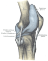
The intercondylar fossa is present between the condyles at the distal end of the femur. In addition to the intercondylar eminence on the tibial plateau, there is both an anterior and posterior intercondylar fossa (area), the sites of anterior cruciate and posterior cruciate ligament attachment, respectively.
In other animals
Parallel structures by the same name exist in other complex animals, such as the bone inside a ham or a leg of lamb. The name femur is also given to the most proximal full-length jointed segment of an arthropod's leg.
References
External links
- Image with major components labeled at v
- Femoral fractures at aofoundation.org
- Cross section image: pembody/body18b—Plastination Laboratory at the Medical University of Vienna
Additional images
-
 Knee diagram
 -
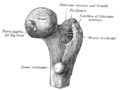 Upper extremity of right femur viewed from behind and above.
 -
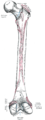
 Right femur. Anterior surface.
 -
 Right femur. Posterior surface.
 -
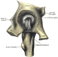 Left hip-joint, opened by removing the floor of the acetabulum from within the pelvis.
 -
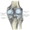
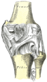 Right knee-joint. Posterior view.
 -
 Left knee-joint from behind, showing interior ligaments.
 -
 Sagittal section of right knee-joint.
 -
 Capsule of right knee-joint (distended). Lateral aspect.
 -
 Capsule of right knee-joint (distended). Posterior aspect.
 -
 Cross-section through the middle of the thigh.
 -
 Deep muscles of the medial femoral region.


