
Glia: Difference between revisions
NeoNeuroGeek (talk | contribs) No edit summary |
|||
| Line 7: | Line 7: | ||
Azevedo FA, Carvalho LR, Grinberg LT, Farfel JM, Ferretti RE, Leite RE, Jacob Filho W, Lent R, Herculano-Houzel S. (2009). Equal numbers of neuronal and nonneuronal cells make the human brain an isometrically scaled-up primate brain. J Comp Neurol. 513(5):532-41. {{PMID|19226510}}</ref> |
Azevedo FA, Carvalho LR, Grinberg LT, Farfel JM, Ferretti RE, Leite RE, Jacob Filho W, Lent R, Herculano-Houzel S. (2009). Equal numbers of neuronal and nonneuronal cells make the human brain an isometrically scaled-up primate brain. J Comp Neurol. 513(5):532-41. {{PMID|19226510}}</ref> |
||
| ⚫ | As the Greek name |
||
==Functions== |
==Functions== |
||
| ⚫ | As the Greek name indicates, glia are commonly known as the [[glue]] or "bricks and mortar" of the nervous system; this, however, is not fully accurate. The four main functions of glial cells are to surround and support neurons by holding them in place, supplying [[nutrients]] and [[oxygen]] to neurons, to insulate one neuron from another, to destroy [[pathogens]], and remove dead neurons. They also modulate [[neurotransmission]].<ref name="gliotransmission">FEBS J. 2008 Jul;275(14):3514-26.d- [http://www.ncbi.nlm.nih.gov/pubmed/18564180 Amino acids in the brain: d-serine in neurotransmission and neurodegeneration.] Wolosker H, Dumin E, Balan L, Foltyn VN.</ref> |
||
While some glial cells function primarily as the physical support for neurons, others nutrify neurons and regulate the [[milieu interieur|internal environment]] of the brain, especially the fluid surrounding neurons and their synapses. During early embryogeny, glial cells direct the migration of neurons and produce molecules that modify the growth of [[axon]]s and [[dendrite]]s. Recent research indicates that glial cells of the [[hippocampus]] and [[cerebellum]] participate in [[synapse|synaptic transmission]], regulate the clearance of neurotransmitters from the synaptic cleft, release factors such as [[adenosine triphosphate|ATP]], which modulate presynaptic function, and even release neurotransmitters themselves. |
|||
Unlike the neuron, which is, in general, considered permanently [[mitosis|post-mitotic]]<ref>Nature Reviews Neuroscience 8, 368-378 (May 2007) | {{doi|10.1038/nrn2124}}</ref>, glial cells are capable of [[mitosis]]. |
Unlike the neuron, which is, in general, considered permanently [[mitosis|post-mitotic]]<ref>Nature Reviews Neuroscience 8, 368-378 (May 2007) | {{doi|10.1038/nrn2124}}</ref>, glial cells are capable of [[mitosis]]. |
||
Revision as of 00:44, 6 September 2010
This article needs additional citations for verification. (February 2008) |


.jpg)
Glial cells, commonly called neuroglia or simply glia (Greek for "glue"), are non-neuronal cells that maintain homeostasis, form myelin, and provide support and protection for the brain's neurons. In the human brain, there is roughly one glia for every neuron with a ratio of about two neurons for every three glia in the cerebral gray matter.[1]
Functions
As the Greek name indicates, glia are commonly known as the glue or "bricks and mortar" of the nervous system; this, however, is not fully accurate. The four main functions of glial cells are to surround and support neurons by holding them in place, supplying nutrients and oxygen to neurons, to insulate one neuron from another, to destroy pathogens, and remove dead neurons. They also modulate neurotransmission.[2] While some glial cells function primarily as the physical support for neurons, others nutrify neurons and regulate the internal environment of the brain, especially the fluid surrounding neurons and their synapses. During early embryogeny, glial cells direct the migration of neurons and produce molecules that modify the growth of axons and dendrites. Recent research indicates that glial cells of the hippocampus and cerebellum participate in synaptic transmission, regulate the clearance of neurotransmitters from the synaptic cleft, release factors such as ATP, which modulate presynaptic function, and even release neurotransmitters themselves. Unlike the neuron, which is, in general, considered permanently post-mitotic[3], glial cells are capable of mitosis.
In the past, glia had been considered to lack certain features of neurons. For example, glial cells were not believed to have chemical synapses or to release neurotransmitters. They were considered to be the passive bystanders of neural transmission. However, recent studies have shown this to be untrue[4]. For example, astrocytes are crucial in clearance of neurotransmitter from within the synaptic cleft, which provides distinction between arrival of action potentials and prevents toxic build-up of certain neurotransmitters such as glutamate (excitotoxicity). It is also thought that glia play a role in Alzheimer's disease. Furthermore, at least in vitro, astrocytes can release neurotransmitter glutamate in response to certain stimulation. Another unique type of glial cell, the oligodendrocyte precursor cells or OPCs, have very well-defined and functional synapses from at least two major groups of neurons. The only notable differences between neurons and glial cells are neurons' possession of axons and dendrites, and capacity to generate action potentials.
Glia are also crucial in the development of the nervous system and in processes such as synaptic plasticity and synaptogenesis. These cells have a role in the regulation of repair of neurons after injury. In the CNS, glia suppresses repair. Glial cells known as astrocytes enlarge and proliferate to form a scar and produce inhibitory molecules that inhibit regrowth of a damaged or severed axon. In the PNS, glial cells known as Schwann cells promote repair. After axonal injury, Schwann cells regress to an earlier developmental state to encourage regrowth of the axon. This difference between PNS and CNS raises hopes for the regeneration of nervous tissue in the CNS. For example, the spinal cord may be able to be repaired following injury or severance.
Types
Microglia
Microglia are like specialized macrophages capable of phagocytosis that protect neurons of the central nervous system. They are derived from hematopoietic precursors rather than ectodermal tissue; they are commonly categorized as such because of their supportive role to neurons.
These cells comprise approximately 15% of the total cells of the central nervous system. They are found in all regions of the brain and spinal cord. Microglial cells are small relative to macroglial cells, with changing shapes and oblong nuclei. They are mobile within the brain and multiply when the brain is damaged. In the healthy central nervous system, microglia processes constantly sample all aspects of their environment (neurons, macroglia and blood vessels).
Macroglia
| Location | Name | Description |
| CNS | Astrocytes |
The most abundant type of macroglial cell, astrocytes (also called astroglia) have numerous projections that anchor neurons to their blood supply. They regulate the external chemical environment of neurons by removing excess ions, notably potassium, and recycling neurotransmitters released during synaptic transmission. The current theory suggests that astrocytes may be the predominant "building blocks" of the blood-brain barrier. Astrocytes may regulate vasoconstriction and vasodilation by producing substances such as arachidonic acid, whose metabolites are vasoactive. Astrocytes signal each other using calcium. The gap junctions (also known as electrical synapses) between astrocytes allow the messenger molecule IP3 to diffuse from one astrocyte to another. IP3 activates calcium channels on cellular organelles, releasing calcium into the cytoplasm. This calcium may stimulate the production of more IP3. The net effect is a calcium wave that propagates from cell to cell. Extracellular release of ATP, and consequent activation of purinergic receptors on other astrocytes, may also mediate calcium waves in some cases. In general, there are two types of astrocytes, protoplasmic and fibrous, similar in function but distinct in morphology and distribution. Protoplasmic astrocytes have short, thick, highly branched processes and are typically found in gray matter. Fibrous astrocytes have long, thin, less branched processes and are more commonly found in white matter. It has recently been shown that astrocyte activity is linked to blood flow in the brain, and that this is what is actually being measured in fMRI. [5] |
| CNS | Oligodendrocytes |
Oligodendrocytes are cells that coat axons in the central nervous system (CNS) with their cell membrane forming a specialized membrane differentiation called myelin, producing the so-called myelin sheath. The myelin sheath provides insulation to the axon that allows electrical signals to propagate more efficiently[6]. |
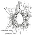
| CNS | Ependymal cells |
Ependymal cells, also named ependymocytes, line the cavities of the CNS and make up the walls of the ventricles. These cells create and secrete cerebrospinal fluid(CSF) and beat their cilia to help circulate that CSF and make up the Blood-CSF barrier. They are also thought to act as neural stem cells[7]. |
| CNS | Radial glia |
Radial glia cells arise from neuroepithelial cells after the onset of neurogenesis. Their differentiation abilities are more restricted than those of neuroepithelial cells. In the developing nervous system, radial glia function both as neuronal progenitors and as a scaffold upon which newborn neurons migrate. In the mature brain, the cerebellum and retina retain characteristic radial glial cells. In the cerebellum, these are Bergmann glia, which regulate synaptic plasticity. In the retina, the radial Müller cell is the principal glial cell, and participates in a bidirectional communication with neurons.[8] |
| PNS | Schwann cells |
Similar in function to oligodendrocytes, Schwann cells provide myelination to axons in the peripheral nervous system (PNS). They also have phagocytotic activity and clear cellular debris that allows for regrowth of PNS neurons.[9] |
| PNS | Satellite cells |
Satellite glial cells are small cells that surround neurons in sensory, sympathetic and parasympathetic ganglia[10]. These cells help regulate the external chemical environment. Like astrocytes, they are interconnected by gap junctions and respond to ATP by elevating intracellular concentration of calcium ions. They are highly sensitive to injury and inflammation, and appear to contribute to pathological states, such as chronic pain. [11] |
| PNS | Enteric glial cells |
Are found in the intrinsic ganglia of the digestive system.They are thought to have many roles in the enteric system, some related to homeostasis and muscular digestive processes.[12] |
Capacity to divide
Glia retain the ability to undergo cell division in adulthood, whereas most neurons cannot. The view is based on the general deficiency of the mature nervous system in replacing neurons after an injury, such as a stroke or trauma, while very often there is a profound proliferation of glia, or gliosis near or at the site of damage. However, detailed studies found no evidence that 'mature' glia, such as astrocytes or oligodendrocytes, retain the ability of mitosis. Only the resident oligodendrocyte precursor cells seem to keep this ability after the nervous system matures. On the other hand, there are a few regions in the mature nervous system, such as the dentate gyrus of the hippocampus and the subventricular zone, where generation of new neurons can be observed.
Embryonic development
Most glia are derived from ectodermal tissue of the developing embryo, in particular the neural tube and crest. The exception is microglia, which are derived from hemopoietic stem cells. In the adult, microglia are largely a self-renewing population and are distinct from macrophages and monocytes, which infiltrate the injured and diseased CNS.
In the central nervous system, glia develop from the ventricular zone of the neural tube. These glia include the oligodendrocytes, ependymal cells, and astrocytes. In the peripheral nervous system, glia derive from the neural crest. These PNS glia include Schwann cells in nerves and satellite glial cells in ganglia.
History
Glia were discovered in 1856 by the pathologist Rudolf Virchow in his search for a 'connective tissue' in the brain.
Numbers
The human brain contains roughly equal numbers of glial cells and neurons with 84.6 billion glia and 86.1 billion neurons.[1] The ratio differs between its different parts. The glia/neuron ratio in the cerebral cortex is 3.72(60.84 billion glia; 16.34 billion neurons) while that of the cerebellum is only 0.23 (16.04 billion glia; 69.03 billion neurons). The ratio in the cerebral cortex gray matter is 1.48 (the white matter part has few neurons). The ratio of the basal ganglia, diencephalon and brainstem combined is 11.35.[1]
Most cerebral cortex glia are oligodendrocytes (75.6%) then astrocytes (17.3%) and least for microglia (6.5%)[13]
The amount of brain tissue that is made up of glia cells increases with brain size: the nematode brain contains only a few glia, a fruitfly's brain is 25% glia; that of a mouse, 65%; a human, 90%; and an elephant, 97%.[14]
Additional images
-
Oligodendrocyte
-
 Section of central canal of medulla spinalis, showing ependymal and neuroglial cells.
 -
 Transverse section of a cerebellar folium.


References
- ^ a b c Azevedo FA, Carvalho LR, Grinberg LT, Farfel JM, Ferretti RE, Leite RE, Jacob Filho W, Lent R, Herculano-Houzel S. (2009). Equal numbers of neuronal and nonneuronal cells make the human brain an isometrically scaled-up primate brain. J Comp Neurol. 513(5):532-41. PMID 19226510
- ^ FEBS J. 2008 Jul;275(14):3514-26.d- Amino acids in the brain: d-serine in neurotransmission and neurodegeneration. Wolosker H, Dumin E, Balan L, Foltyn VN.
- ^ Nature Reviews Neuroscience 8, 368-378 (May 2007) | doi:10.1038/nrn2124
- ^ The Other Brain, by R. Douglas Fields, Ph. D. Simon & Schuster, 2009
- ^ Swaminathan N (2008). "Brain-scan mystery solved". Scientific American Mind. Oct–Nov: 7.
- ^ Baumann, Nicole; Pham-Dinh, Danielle (2001), "Biology of Oligodendrocyte and Myelin in the Mammalian Central Nervous System", Physiological Reviews 18 (2): 871–927
- ^ Johansson CB, Momma S, Clarke DL, Risling M, Lendahl U, Frisen J (1999). "Identification of a neural stem cell in the adult mammalian central nervous system". Cell 96 (1): 25–34
- ^ Campbell K, Götz M (May 2002). "Radial glia: multi-purpose cells for vertebrate brain development". Trends Neurosci. 25 (5): 235–8.
- ^ Jessen, K. R. & Mirsky, R. (2005), "The origin and development of glial cells in peripheral nerves", Nature Reviews Neuroscience 6 (9): 671–682
- ^ Hanani, M. Satellite glial cells in sensory ganglia: from form to function. Brain Res. Rev. 48:457-476, 2005
- ^ Ohara PT et al., Evidence for a role of connexin 43 in trigeminal pain using RNA interference in vivo. J Neurophysiol 2008;100:3064-3073
- ^ Bassotti, G. et al, Laboratory Investigation (2007) 87, 628–632
- ^ Pelvig DP, Pakkenberg H, Stark AK, Pakkenberg B. (2008). Neocortical glial cell numbers in human brains. Neurobiol Aging. 29(11):1754-62.(figures given are those for females) PMID 17544173
- ^ Allen NJ, Barres BA. (2009). Neuroscience: Glia - more than just brain glue. Nature. 457(7230):675-7. PMID 19194443
External links
 Media related to Glia at Wikimedia Commons
- "The Mystery and Magic of Glia: A Perspective on Their Roles in Health and Disease." Neuron 60, November 6, 2008 by Ben Barres
- Role of glia in synapse development
- Overstreet L (2005). "Quantal transmission: not just for neurons". Trends Neurosci. 28 (2): 59–62. doi:10.1016/j.tins.2004.11.010. PMID 15667925. article
- Peters A (2004). "A fourth type of neuroglial cell in the adult central nervous system". J Neurocytol. 33 (3): 345–57. doi:10.1023/B:NEUR.0000044195.64009.27. PMID 15475689.
- Volterra A, Steinhäuser C (2004). "Glial modulation of synaptic transmission in the hippocampus". Glia. 47 (3): 249–57. doi:10.1002/glia.20080. PMID 15252814.
- Huang Y, Bergles D (2004). "Glutamate transporters bring competition to the synapse". Curr Opin Neurobiol. 14 (3): 346–52. doi:10.1016/j.conb.2004.05.007. PMID 15194115.
- New Source of Replacement Brain Cells Found - glial cells can transform into other cell types and reproduce indefinitely — tricks once thought exclusive to stem cells.
- Artist ADSkyler(uses concepts of neuroscience and found inspiration from Glia)
- Futures in Biotech Episode 63 - Interview with Dr. Mario Capecchi discussing (among other things) his work on the role of glia in OCD.