
Carpal tunnel: Difference between revisions
m →Structure: hammate spelling corrected to hamate |
Add correct section anchor to link |
||
| Line 20: | Line 20: | ||
DorlandsSuf = 12208551 | |
DorlandsSuf = 12208551 | |
||
}} |
}} |
||
In the [[human body]], the '''carpal tunnel''' or '''carpal canal''' is the passageway on the [[Anatomical terms of location|palmar]] side of the [[wrist]] that connects the forearm to the middle compartment of the deep plane of the palm.<ref name="Schmidt-29">{{cite book |
In the [[human body]], the '''carpal tunnel''' or '''carpal canal''' is the passageway on the [[Anatomical terms of location#Arms|palmar]] side of the [[wrist]] that connects the forearm to the middle compartment of the deep plane of the palm.<ref name="Schmidt-29">{{cite book |
||
| title = Surgical anatomy of the hand |
| title = Surgical anatomy of the hand |
||
| url = http://books.google.com/books?id=L7a1tkBU8eMC&pg=PA29 |
| url = http://books.google.com/books?id=L7a1tkBU8eMC&pg=PA29 |
||
Revision as of 23:03, 30 August 2013
| Carpal tunnel | |
|---|---|
 | |
| Details | |
| Identifiers | |
| Latin | Canalis carpi |
| TA98 | A03.5.11.201 |
| TA2 | 2551 |
| FMA | 42352 |
| Anatomical terminology | |
In the human body, the carpal tunnel or carpal canal is the passageway on the palmar side of the wrist that connects the forearm to the middle compartment of the deep plane of the palm.[1] The tunnel consists of bones and connective tissue. Several tendons and a nerve pass through it.
The canal is narrow and when any of the nine long flexor tendons passing through it swells or degenerates, the narrowing of the canal often results in the median nerve becoming entrapped or compressed, a medical condition known as carpal tunnel syndrome.[2]
Contents
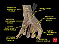
A total of nine flexor tendons[2] (not the muscles themselves) pass through the carpal tunnel:
- flexor digitorum profundus (four tendons)
- flexor digitorum superficialis (four tendons)
- flexor pollicis longus (one tendon)
A single nerve passes through the tunnel: the median nerve between tendons of flexor digitorum profundus and flexor digitorum superficialis
Structure
The carpus, the bony elements of the wrist, form an arch which is convex on the dorsal side of the hand and concave on the palmar side. The groove on the palmar side, the sulcus carpi, is covered by the flexor retinaculum, a sheath of tough connective tissue, thus forming the carpal tunnel. The flexor retinaculum is attached radially to the scaphoid tubercle and the ridge of trapezium, and on the ulna side to the pisiform and hook of hamate.[3]
The narrowest section of the tunnel is located a centimetre beyond the mid-line of the distal row of carpal bones where the sectional area is limited to 1.6 cm2.[2]
The tendons of the flexor digitorum superficialis and profundus pass through a common ulnar sheath, while the tendon of the flexor pollicis longus passes through a separate radial sheath. The mesotendon shared by these tendons is attached to the radial and palmar walls of the carpal tunnel.[3]
Superficial to the carpal tunnel and the flexor retinaculum, the ulnar artery and ulnar nerve pass through the ulnar tunnel.[3]
Effect of wrist movements
Movements in the wrist affect the shape and width of the carpal tunnel. The width decreases considerably during normal range of motion in the wrist and because the carpal bones move in relation to each other with every motion of the hand the bony walls of the tunnel are not rigid. Both flexion and extension increase compression in the carpal tunnel.
- Flexing the wrist causes the flexor retinaculum to move closer to the radius which considerably decreases the cross section of the proximal opening of the tunnel. Additionally, the distal end of the capitate presses into the opening.
- In extreme extension, the lunate constricts the passage as it is pressed toward the interior of the tunnel.[1]
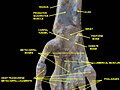
Additional Images
-
 Carpal tunnel
 -
 Carpal tunnel
 -
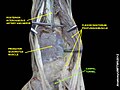
 Wrist joint. Deep dissection.Anterior, palmar, view.
 -
 Wrist joint. Deep dissection.Anterior, palmar, view.
 -
 Wrist joint. Deep dissection.Anterior, palmar, view.
 -
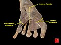 Carpal tunnel and thenar and hypothenar eminences
 -
 Carpal tunnel


See also
References
- ^ a b Schmidt, Hans-Martin; Lanz, Ulrich (2003). Surgical anatomy of the hand. Thieme. p. 29. ISBN 1-58890-007-X.
- ^ a b c Thieme Atlas of Anatomy: General Anatomy and Musculoskeletal System. Thieme. 2006. pp. 248–249. ISBN 1-58890-419-9.
- ^ a b c Thieme Atlas of Anatomy: General Anatomy and Musculoskeletal System. Thieme. 2006. p. 354. ISBN 1-58890-419-9.