Cranial nerve nucleus: Difference between revisions
Appearance
Content deleted Content added
m Replace magic links with templates per local RfC and MediaWiki RfC |
Rescuing 1 sources and tagging 0 as dead. #IABot (v1.5beta) |
||
| Line 104: | Line 104: | ||
==External links== |
==External links== |
||
* {{UMichAtlas|n2a6p2}} |
* {{UMichAtlas|n2a6p2}} |
||
* [http://www.coloradocollege.edu/idprog/Neuroscience/Cranial.html Slides at Colorado College] |
* [https://web.archive.org/web/20060113143844/http://www.coloradocollege.edu/idprog/Neuroscience/Cranial.html Slides at Colorado College] |
||
{{Cranial nerves}} |
{{Cranial nerves}} |
||
Revision as of 05:00, 14 August 2017
| Cranial nerve nucleus | |
|---|---|
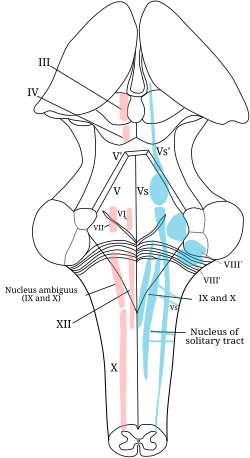
 The cranial nerve nuclei schematically represented; dorsal view. Motor nuclei in red; sensory in blue. (The olfactory and optic centers are not represented.) | |
| Details | |
| Identifiers | |
| Latin | nucleus nervi cranialis |
| NeuroLex ID | nlx_28532 |
| TA98 | A14.1.00.004 |
| TA2 | 5857 |
| FMA | 54501 |
| Anatomical terms of neuroanatomy | |
A cranial nerve nucleus is a collection of neurons (gray matter) in the brain stem that is associated with one or more cranial nerves. Axons carrying information to and from the cranial nerves form a synapse first at these nuclei. Lesions occurring at these nuclei can lead to effects resembling those seen by the severing of nerve(s) they are associated with. All the nuclei except that of the trochlear nerve (CN IV) supply nerves of the same side of the body.
Structure
Motor and sensory
In general, motor nuclei are closer to the front (ventral), and sensory nuclei and neurons are closer to the back (dorsal). This arrangement mirrors the arrangement of tracts in the spinal cord.
- Close to the midline are the motor efferent nuclei, such as the oculomotor nucleus, which control skeletal muscle. Just lateral to this are the autonomic (or visceral) efferent nuclei.
- There is a separation, called the sulcus limitans, and lateral to this are the sensory nuclei. Near the sulcus limitans are the visceral afferent nuclei, namely the solitary tract nucleus.
- More lateral, but also less posterior, are the general somatic afferent nuclei. This is the trigeminal nucleus. Back at the dorsal surface of the brainstem, and more lateral are the special somatic afferents, this handles sensation such as balance.
- Another area, not on the dorsum of the brainstem, is where the special visceral efferents nuclei reside. These formed from the pharyngeal arches, in the embryo. This area is a bit below the autonomic motor nuclei, and includes the nucleus ambiguus, facial nerve nucleus, as well as the motor part of the trigeminal nerve nucleus.
Location
This list documents nuclei by the part of the brain they are found in:
Nuclei present in the Midbrain
- Mesencephalic trigeminal nucleus (V) - sensory
- Red nucleus - motor, extrapyramidal
- Trochlear nucleus (IV) - motor
- Oculomotor nucleus (III) - motor
- Edinger-Westphal nucleus (III) - visceromotor
Nuclei present in the Pons
- Cochlear nuclei (VIII) - sensory
- Vestibular nuclei (VIII) - sensory
- Salivary nuclei - visceromotor
- Facial nucleus (VII) - motor
- Abducens nucleus (VI) - motor
- Trigeminal motor nucleus (V) - motor
- Main trigeminal nucleus (V) - sensory (fine touch and vibration)
Nuclei present in the medulla

- Hypoglossal nucleus (XII) - motor
- Dorsal motor nucleus of vagus nerve (X) - visceromotor
- Nucleus ambiguus (IX, X, XI) - motor
- Solitary nucleus (VII, IX, X) - sensory
- Spinal trigeminal nucleus (V) - sensory (crude touch, temperature and pain)
- Inferior olivary nucleus afferent fibres to cerebellum
Location
| Olfactory nerve | Olfactory bulb |
| Optic nerve | Lateral geniculate nucleus |
| Oculomotor nerve | Oculomotor nucleus Edinger-Westphal nucleus |
| Trochlear nerve | Trochlear nucleus |
| Trigeminal nerve | Trigeminal nerve nuclei: Mesencephalic nucleus Principal sensory nucleus Spinal trigeminal nucleus Trigeminal motor nucleus |
| Abducens nerve | Abducens nucleus |
| Facial nerve | Facial motor nucleus Superior salivatory nucleus Solitary nucleus |
| Vestibulocochlear nerve | Vestibular nuclei w. subnuclei Cochlear nucleus w. subnuclei |
| Glossopharyngeal nerve | Solitary nucleus Spinal nucleus of the trigeminal nerve Lateral nucleus of vagal trigone. Nucleus ambiguus Inferior salivatory nucleus |
| Vagus nerve | Dorsal nucleus of vagus nerve Nucleus ambiguus Solitary nucleus Spinal trigeminal nucleus |
| Accessory nerve | Spinal accessory nucleus Nucleus ambiguus |
| Hypoglossal nerve | Hypoglossal nucleus |
References
- Lennart Heimer, The Human Brain, ISBN 0-387-94227-0
Additional images
-
Nuclei of origin of cranial motor nerves schematically represented; lateral view.
-
Primary terminal nuclei of the afferent (sensory) cranial nerves schematically represented; lateral view.
-
Brain stem sagittal section
External links
- Atlas image: n2a6p2 at the University of Michigan Health System
- Slides at Colorado College


