
Facial colliculus: Difference between revisions
Content deleted Content added
m cleanup (wikitables, html markup, layout, etc.) |
Rescuing 1 sources and tagging 0 as dead. #IABot (v1.5.4) |
||
| Line 31: | Line 31: | ||
* http://www.med.yale.edu/caim/cnerves/cn6/cn6_2.html |
* http://www.med.yale.edu/caim/cnerves/cn6/cn6_2.html |
||
* http://www.neuroanatomy.wisc.edu/virtualbrain/BrainStem/14CNVII.html |
* http://www.neuroanatomy.wisc.edu/virtualbrain/BrainStem/14CNVII.html |
||
* http://www.ib.amwaw.edu.pl/anatomy/atlas/image_04be.htm |
* https://web.archive.org/web/20070927162218/http://www.ib.amwaw.edu.pl/anatomy/atlas/image_04be.htm |
||
{{Pons}} |
{{Pons}} |
||
Revision as of 10:45, 27 September 2017
| Facial colliculus | |
|---|---|
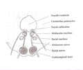
 Rhomboid fossa. (Colliculus facialis labeled at center left.) | |
 Human caudal brainstem posterior view (Colliculus facialis is #3) | |
| Details | |
| Identifiers | |
| Latin | colliculus facialis |
| NeuroNames | 624 |
| TA98 | A14.1.05.705 |
| FMA | 78480 |
| Anatomical terms of neuroanatomy | |
The facial colliculus is an elevated area located on the dorsal pons in the floor of the 4th ventricle. It is formed by fibers from the motor nucleus of the facial nerve as they loop over the abducens nucleus. Thus a lesion to the facial colliculus would result in ipsilateral facial paralysis and ipsilateral unopposed eye medial deviation.
Additional images
-
 Axial section of the Brainstem (Pons) at the level of the Facial Colliculus
 -
 Fourth ventricle. Posterior view.Deep dissection.


External links
- http://www.med.yale.edu/caim/cnerves/cn6/cn6_2.html
- http://www.neuroanatomy.wisc.edu/virtualbrain/BrainStem/14CNVII.html
- https://web.archive.org/web/20070927162218/http://www.ib.amwaw.edu.pl/anatomy/atlas/image_04be.htm
This neuroanatomy article is a stub. You can help Wikipedia by expanding it. |