Buccopharyngeal fascia
| Buccopharyngeal fascia | |
|---|---|
 Carotid sheath outlined in red | |
 | |
| Details | |
| Identifiers | |
| Latin | fascia buccopharyngea |
| TA98 | A04.1.04.010 A05.3.01.116 |
| TA2 | 2211 |
| FMA | 55078 |
| Anatomical terminology | |
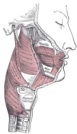
The buccopharyngeal fascia is a fascia of the pharynx.[1] It represents the posterior portion of the pretracheal fascia[2] (visceral fascia).[3] It covers the superior pharyngeal constrictor muscles, and buccinator muscle.[4]
Structure
[edit]The buccopharyngeal fascia is a thin lamina given off from the pretracheal fascia.[citation needed] It is the portion of the pretracheal fascia situated posterior and lateral to the pharynx. It encloses the entire superior part of the alimentary canal.[3]
The buccopharyngeal fascia envelops the superior pharyngeal constrictor muscles.[4][1] It extends anteriorly from the constrictor pharyngis superior[4] over the pterygomandibular raphe to cover the buccinator muscle[1] (though another source describes it as continuous with the fascia covering the buccinator muscle).[3]
Attachments
[edit]It is attached to the prevertebral fascia by loose connective tissue, with the retropharyngeal space found between them.[citation needed] It may also be attached to the alar fascia posteriorly at C3 and C6 levels.[5]
Relations
[edit]The thyroid gland wraps around the trachea and oesophagus anterior to the buccopharyngeal fascia, so that the lateral parts of the thyroid gland border it.[6]
The buccopharyngeal fascia runs parallel to the medial aspect of the carotid sheath.[citation needed]
Additional images
[edit]-
Floor of mouth. Deep dissection.Anterior view.
See also
[edit]References
[edit]![]() This article incorporates text in the public domain from page 390 of the 20th edition of Gray's Anatomy (1918)
This article incorporates text in the public domain from page 390 of the 20th edition of Gray's Anatomy (1918)
- ^ a b c Standring, Susan (2020). Gray's Anatomy: The Anatomical Basis of Clinical Practice (42nd ed.). New York. p. 709. ISBN 978-0-7020-7707-4. OCLC 1201341621.
{{cite book}}: CS1 maint: location missing publisher (link) - ^ Morton, David A. (2019). The Big Picture: Gross Anatomy. K. Bo Foreman, Kurt H. Albertine (2nd ed.). New York. p. 266. ISBN 978-1-259-86264-9. OCLC 1044772257.
{{cite book}}: CS1 maint: location missing publisher (link) - ^ a b c Fehrenbach, Margaret J.; Herring, Susan W. (2017). Illustrated Anatomy of the Head and Neck (5th ed.). St. Louis: Elsevier. pp. 266–267. ISBN 978-0-323-39634-9.
- ^ a b c Stecco, Carla; Hammer, Warren; Vleeming, Andry; De Caro, Raffaele (2015-01-01), Stecco, Carla; Hammer, Warren; Vleeming, Andry; De Caro, Raffaele (eds.), "4 - Fasciae of the Head and Neck", Functional Atlas of the Human Fascial System, Churchill Livingstone, pp. 103–139, ISBN 978-0-7020-4430-4, retrieved 2021-01-12
- ^ Yousem, David M. (2015-01-01), Yousem, David M. (ed.), "Fair Game", Head and Neck Imaging (Fourth Edition), Case Review Series, Philadelphia: W.B. Saunders, pp. 103–304, ISBN 978-1-4557-7629-0, retrieved 2021-01-13
- ^ Thompson, Stevan H.; Yeung, Alison Y. (2016-01-01), Hupp, James R.; Ferneini, Elie M. (eds.), "4 - Anatomy Relevant to Head, Neck, and Orofacial Infections", Head, Neck, and Orofacial Infections, St. Louis: Elsevier, pp. 60–93, doi:10.1016/b978-0-323-28945-0.00004-1, ISBN 978-0-323-28945-0, retrieved 2021-01-13
External links
[edit]- Anatomy photo:31:10-0102 at the SUNY Downstate Medical Center - "Pharynx: The Pharyngeal Constrictor Muscles"
- "Buccopharyngeal fascia". Medcyclopaedia. GE. Archived from the original on 2012-02-05.

