
Epiglottis
| Epiglottis | |
|---|---|
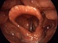
 Posterior view of the larynx. The epiglottis is the most superior structure shown. | |
| Details | |
| Precursor | Hypobranchial eminence[1][unreliable source?] |
| Identifiers | |
| MeSH | D004825 |
| TA98 | A06.2.07.001 |
| TA2 | 3190 |
| FMA | 55130 |
| Anatomical terminology | |
The epiglottis is a flap of elastic cartilage tissue covered with a mucus membrane, attached to the root of the tongue. It projects obliquely upwards behind the tongue and the hyoid bone, pointing dorsally. The term is, like tonsils, often incorrectly used to refer to the uvula.[2]
Anatomy and function
The epiglottis guards the entrance of the glottis, the opening between the vocal folds. It is normally pointed upward during breathing with its underside functioning as part of the pharynx, but during swallowing, elevation of the hyoid bone draws the larynx upward; as a result, the epiglottis folds down to a more horizontal position, with its upper side functioning as part of the pharynx. In this manner it prevents food from going into the trachea and instead directs it to the esophagus, which is posterior.
The epiglottis is one of nine cartilaginous structures that make up the larynx (voice box). While breathing, it lies completely within the pharynx. When swallowing it serves as part of the anterior of the larynx.[citation needed]
Clinical significance
Reflexes
The glossopharyngeal nerve (CN IX) sends fibers to the upper epiglottis that contribute to the afferent limb of the gag reflex. The superior laryngeal branch of the vagus nerve (CN X) sends fibers to the lower epiglottis that contribute to the efferent limb of the cough reflex.[3]
Infection of the epiglottis
In children, the epiglottis will occasionally become infected with Haemophilus influenzae and Streptococci in the trachea, causing massive inflammation, called "Epiglottitis". This condition has become rare in countries where vaccination against Haemophilus influenzae (Hib) is administered.[citation needed]
Additional images
-
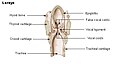 Larynx
 -
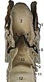 Cut through the larynx of a horse
 -
 The cartilages of the larynx. Posterior view.
 -
 Ligaments of the larynx. Posterior view.
 -
 Coronal section of larynx and upper part of trachea.
 -
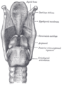
 The entrance to the larynx, viewed from behind.
 -
 Muscles of larynx. Posterior view.
 -
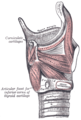 Muscles of larynx. Side view. Right lamina of thyroid cartilage removed.
 -
 Sagittal section of nose mouth, pharynx, and larynx.
 -
 Larynx. 1=vocal cords, 2=vestibular fold, 3=epiglottis, 4=plica aryepiglottica, 5=arytenoid cartilage, 6=sinus piriformis, 7=dorsum of the tongue


References
- ^ Stevenson, Roger E. (2006). Human malformations and related anomalies. Oxford [Oxfordshire]: Oxford University Press. ISBN 0-19-516568-3.
- ^ http://www.thefirstpost.co.uk/45548,features,someones-been-careless-with-my-eplglottis
- ^ April, Ernest. Clinical Anatomy, 3rd ed. Lippincott, Williams, and Wilkins.
External links
- lesson11 at The Anatomy Lesson by Wesley Norman (Georgetown University) (larynxsagsect)