Microscope

A microscope (Greek: micron = small and scopos = aim) is an instrument for viewing objects that are too small to be seen by the naked or unaided eye. The science of investigating small objects using such an instrument is called microscopy, and the term microscopic means minute or very small, not easily visible with the unaided eye. In other words, requiring a microscope to examine.
The most common type of microscope—and the first to be invented—is the optical microscope. This is an optical instrument containing one or more lenses that produce an enlarged image of an object placed in the focal plane of the lens(es).
See also: Microscopy.
Simple optical microscope
A simple microscope, as opposed to a standard compound microscope (see below) with multiple lenses, is a microscope that uses only one lens for magnification. Van Leeuwenhoek's microscopes consisted of a single, small, convex lens mounted on a plate with a mechanism to hold the material to be examined (the sample or specimen). This use of a single, convex lens to magnify objects for viewing is still found in the magnifying glass, the hand-lens, and the loupe.
Compound optical microscope
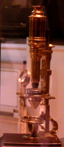
The diagram below shows a compound microscope. In its simplest form—as used by Robert Hooke, for example—the compound microscope would have a single glass lens of short focal length for the objective, and another single glass lens for the eyepiece or ocular. Modern microscopes of this kind are usually more complex, with multiple lens components in both objective and eyepiece assemblies. These multi-component lenses are designed to reduce aberrations, particularly chromatic aberration and spherical aberration. In modern microscopes the mirror is replaced by a lamp unit providing stable, controllable illumination.
|
The Parts of the Microscope
There are eleven main parts to a microscope. At the top of the microscope there is a cylinder that is called the ocular or eyepiece. That is where you look to see the magnified image. The ocular is connected to the body tube that leads to the main microscope. The hanging cylinders are called the objective lenses and can have many magnifications. They are attached to the nose piece. Right under them is the stage where the item is placed and a diaphragm is attached under the stage to control how much light or what color you view the object with. Right under the diaphragm is the light source. Next to the light source, there are two knobs. The small knob is the fine adjustment and the big knob is the coarse adjustment. These knobs are usually located on the arm. Last but not least, if the microscope is electronic there should be a power switch.
Compound optical microscopes can magnify an image up to 1000× and are used to study thin specimens as they have a very limited depth of field. Typically they are used to examine a smear, a squash preparation, or a thinly sectioned slice of some material. With a few exceptions, they utilize light passing through the sample from below and special techniques are usually necessary to increase the contrast in the image to useful levels (see contrast methods). Typically, on a standard compound optical microscope, there are three objective lenses: a scanning lens (4×), low power lens (10×), and high power lens (40×). Advanced microscopes often have a fourth objective lens, called an oil immersion lens. To use this lens, a drop of oil is placed on top of the cover slip, and the lens moved into place where it is immersed in the oil. An oil immersion lens usually has a power of 100×. The actual power or magnification is the product of the powers of the ocular (eyepiece), usually about 10×, and the objective lens being used.
To study the thin structure of metals (see metallography) and minerals, another type of microscope is used, where the light is reflected from the examined surface. The light is fed through the same objective using a semi-transparent mirror.
How a microscope works
The optical components of a modern microscope are very complex and for a microscope to work well, the whole optical path has to be very accurately set up and controlled. Despite this , the basic optical principles of a microscope are quite simple.
The objective lens is, at its simplest, a very high powered magnifying glass i.e. a lens with a very short focal length. This is brought very close to the specimen being examined so that the light from the specimen comes to a focus about 160 mm inside the microscope tube. This creates a virtual and enlarged image of the subject. This image is inverted and can be seen by removing the eyepiece and placing a piece of tracing paper over the end of the tube. By careful focusing a rather dim image of the specimen, much enlarged can be seen. It is this virtual image that is viewed by the eyepiece lens that provides further enlargement.
In most microscopes, the eyepiece is a compound lens, which is made of two lenses one near the front and one near the back of the eyepiece tube forming an air separated couplet. In many designs, the virtual image comes to a focus between the two lenses of the eyepiece, the first lens bringing the virtual image to a focus and the second lens enabling the eye to focus on the image.
In all microscopes the image is viewed with the eyes focused at infinity. Headaches and tired eyes after using a microscope are usually signs that the eye is being forced to focus at a close distance rather than at infinity.
Stereomicroscope
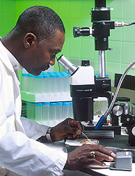
The stereo or dissecting microscope is designed differently from the diagrams above, and serves a different purpose. It uses two separate optical paths with two objectives and two eypieces to provide slightly different viewing angles to the left and right eyes. In this way it produces a three-dimensional (3-D) visualisation of the sample being examined.

The stereo microscope is often used to study the surfaces of solid specimens or to carry out close work such as sorting, dissection, microsurgery, watch-making, small circuit board manufacture or inspection, and the like.
Great working distance and depth of field here are important qualities for this type of microscope. Both qualities are inversely correlated with resolution: the higher the resolution (i.e., magnification), the smaller the depth of field and working distance. A stereo microscope has a useful magnification up to 100×. The resolution is maximally in the order of an average 10× objective in a compound microscope, and often much lower.
The stereo-microscope should not be confused with ordinary compound microscopes equipped with a binocular eyepieces. In these microscopes both eyes can see the image but the binocular head provides greater viewing comfort and slightly better appearance of resolution. However the image in such microscopes remains monocular.
Special designs
Other types of optical microscope include:
- the inverted microscope for studying samples from below; useful for cell cultures in liquid;
- the student microscope designed for low cost, durability, and ease of use; and
- the research microscope which is an expensive tool with many enhancements.
- the petrographic microscope whose design usually includes a polarizing filter, rotating stage and gypsum plate to facilitate the study of minerals or other crystalline materials whose optical properties can vary with orientation.
Optical Resolution
A lens magnifies by bending light (see refraction). Optical microscopes are restricted in their ability to resolve features by a phenomenon called diffraction which, based on the numerical aperture (NA or ) of the optical system and the wavelengths of light used (), sets a definite limit (d) to the optical resolution. Assuming that optical aberrations are negligible, the resolution (d) is given by:
Usually, a of 550 nm is assumed, corresponding to green light. With air as medium, the highest practical is 0.95, and with oil, up to 1.5.
Due to diffraction, even the best optical microscope is limited to a resolution of 0.2 micrometres.
History of the microscope

It is impossible to say who invented the compound microscope. Dutch spectacle-makers Hans Janssen and his son Zacharias Janssen are often said to have invented the first compound microscope in 1590, but this was a declaration by Zacharias Janssen himself halfway through the 17th century. The date is certainly not likely, as it has been shown that Zacharias Janssen actually was just about born in 1590. Another favorite for the title of 'inventor of the microscope' was Galileo Galilei. He developed an occhiolino or compound microscope with a convex and a concave lens in 1609. Galilei´s microscope was celebrated in the ´Lynx academy´ founded by Federico Cesi in 1603. Francesco Stelluti´s drawing of three bees were part of pope Urban VIII´s seal, and count as the first microscopic figure published (see Stephen Jay Gould, The Lying stones of Marrakech, 2000). Christiaan Huygens, another Dutchman, developed a simple 2-lens ocular system in the late 1600's that was achromatically corrected and therefore a huge step forward in microscope development. The Huygens ocular is still being produced to this day, but suffers from a small field size, and the eye relief is uncomfortably close compared to modern widefield oculars.
Anton van Leeuwenhoek (1632-1723) is generally credited with bringing the microscope to the attention of biologists, even though simple magnifying lenses were already being produced in the 1500's, and the magnifying principle of water-filled glass bowls had been described by the Romans (Seneca). Van Leeuwenhoek's home-made microscopes were actually very small simple instruments with a single very strong lens. They were awkard in use but enabled van Leeuwenhoek to see highly detailed images, mainly because a single lens does not suffer the lens faults that are doubled or even multiplied when using several lenses in combination as in a compound microscope. It actually took about 150 years of optical development before the compound microscope was able to provide the same quality image as van Leeuwenhoek's simple microscopes. So although he was certainly a great microscopist, van Leeuwenhoek is, contrary to widespread claims, certainly not the inventor of the microscope.
Other types of microscopes

See also microscopy
- Atom probe
- Atomic force microscope
- Darkfield microscope
- Electron microscope
- Field ion microscope
- Field emission microscope
- Phase contrast microscope, see Frits Zernike
- Scanning tunneling microscope
- Virtual microscope
- X-ray microscope
- Total internal reflection fluorescence microscope
- Confocal laser scanning microscopy
See also
- Angular resolution
- Microscope image processing
- Microscope slide
- Microscopy
- Microscopy laboratory in: A Study Guide to the Science of Botany at Wikibooks
- Telescope
External links
- A virtual polarization microscope (requires Java)
- How To Buy A Microscope
- Micscape - a monthly magazine directed towards the amateur microscopist
- Microscopy
- Microscope Directory
- the optics of the microscope
- Optical microscopy primer
- Royal Microscopical Society
- The Microscope - quarterly journal
- Antique German microscopes history of continental microscopes illustrated with 2000 photos (in German)
- Early American made microscopes Antique American made microscopes and the makers.
- Some Early Microscopes from the Optical Institute in Wetzlar Microscope history.


