
Recurrent laryngeal nerve: Difference between revisions
Novangelis (talk | contribs) Degrees do not establish reliability of sources; Sources fail reliability. |
No edit summary |
||
| (One intermediate revision by the same user not shown) | |||
| Line 58: | Line 58: | ||
==Evidence of evolution== |
==Evidence of evolution== |
||
The extreme detour of this nerve (about 15 feet in the case of [[giraffe]]s<ref>{{Cite book | title=Mammal Anatomy: An Illustrated Guide | year=2010 | publisher=Marshall Cavendish Corporation | pages=74–75 | isbn=0-7614-7882-5 }}</ref>) is cited as [[evidence of evolution]]. The nerve's route would have been direct in the fish-like ancestors of modern tetrapods, traveling from the brain, past the heart, to the gills (as it does in modern fish). Over the course of evolution, as the neck extended and the heart became lower in the body, the laryngeal nerve was caught on the wrong side of the heart. Natural selection gradually lengthened the nerve by tiny increments to accommodate, resulting in the circuitous route now observed.<ref>{{cite book |last= Dawkins |first= Richard |authorlink= Richard Dawkins |title= The greatest show on Earth |url= http://books.google.com/books?id=U8AFxmc76rcC |accessdate= November 21, 2009 |year= 2009 |publisher= Free Press |location= New York |isbn= 978-1-4165-9478-9 |pages= 360–362 |chapter= 11. History written all over us}}</ref> |
The extreme detour of this nerve (about 15 feet in the case of [[giraffe]]s<ref>{{Cite book | title=Mammal Anatomy: An Illustrated Guide | year=2010 | publisher=Marshall Cavendish Corporation | pages=74–75 | isbn=0-7614-7882-5 }}</ref>) is cited as [[evidence of evolution]]. The nerve's route would have been direct in the fish-like ancestors of modern tetrapods, traveling from the brain, past the heart, to the gills (as it does in modern fish). Over the course of evolution, as the neck extended and the heart became lower in the body, the laryngeal nerve was caught on the wrong side of the heart. Natural selection gradually lengthened the nerve by tiny increments to accommodate, resulting in the circuitous route now observed.<ref>{{cite book |last= Dawkins |first= Richard |authorlink= Richard Dawkins |title= The greatest show on Earth |url= http://books.google.com/books?id=U8AFxmc76rcC |accessdate= November 21, 2009 |year= 2009 |publisher= Free Press |location= New York |isbn= 978-1-4165-9478-9 |pages= 360–362 |chapter= 11. History written all over us}}</ref> |
||
On the other side of the debate, the route of the laryngeal nerve is seen as the result of developmental constraints.<ref>{{cite web|last=Lönnig|first=Wolf-Ekkehard|title=The Laryngeal Nerve of the Giraffe: Does it Prove Evolution?|url=http://www.weloennig.de/LaryngealNerve.pdf|publisher=Planck Institute for Plant Breeding Research|location=Germany|pages=3}}</ref><ref>Blechschmidt, E. 2004. The Ontogenetic Basis of Human Anatomy: A Biodynamic Approach to Development from Conception to Birth. B. Freeman, transl. New York: North Atlantic Books, 188. </ref><ref>Bergman, J. 2010. Recurrent Laryngeal Nerve Is Not Evidence of Poor Design. Acts & Facts. 39 (8): 12-14.</ref> When the body begins as a spherical blastocyst and elongates with development, vestigial arteries and other structures are perforce elongated and moved to remain functional throughout growth. The movement and morphology of the nerve could have resulted from the formation of the neck and the body's elongation during the fetal phase, as the heart is forced downwards from the cervical (neck) location into the thoracic (chest) cavity.<ref>Sadler, T. W. 1990. Langman's Medical Embryology, 6th ed. Philadelphia, PA: Williams & Wilkins, 211.</ref> The ''right'' tangent of the nerve could have been carried downward because it's looped under the arch that develops into the ''right'' subclavian artery, thus causing it to move down as development continued.<ref>Schoenwolf, G. C., S. B. Bleyl, P. R. Brauer and P. H. Francis-West. 2009. Larsen's Human Embryology. Philadelphia, PA: Churchill Livingstone, 407.</ref> The ''left'' tangent of the laryngeal nerve that recurs around the ''ligamentum arteriosum'' on the left side of the aortic arch, could have likewise moved down in tandem as the thoracic cavity lengthened. As the result of this downward movement of the heart, the course of the recurrent laryngeal nerves could have changed on each side.<ref>Sadler, 1990, Langman's Medical Embryology, 211.</ref> The nerve also serves several other organs with motor and sensory branches (i.e.: upper esophagus, trachea, inferior pharynx, and the cricopharynxgeus muscle).<ref>Sturniolo et al, The Recurrent Laryngeal Nerve Related to Thyroid Surgery, 487.</ref><ref>{{cite web|last=Gray|first=Henry|title=Gray's Anatomy|url=http://books.google.com/books?id=uaQMAAAAYAAJ&pg=PA912&lpg#v=onepage&q&f=false|pages=912}}</ref> In addition, several studies have discovered that the existing course of the nerve renders it less prone to damage or injury than a more direct route.<ref>Armstrong, W. G. and J. W. Hinton. 1951. Multiple Divisions of the Recurrent Laryngeal Nerve. AMA Archives of Surgery. 62 (4): 539.</ref> |
|||
==References== |
==References== |
||
Revision as of 05:13, 11 August 2013
The article's lead section may need to be rewritten. (June 2012) |
| Recurrent laryngeal nerve | |
|---|---|
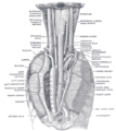
 POSTERIOR VIEW: The tracheobronchial lymph glands (I. and E. Recurrent nerves visible at top.) | |
 | |
| Details | |
| From | vagus nerve |
| Innervates | larynx posterior cricoarytenoid lateral cricoarytenoid arytenoid thyroarytenoid aryepiglottis |
| Identifiers | |
| Latin | nervus laryngeus recurrens |
| MeSH | D012009 |
| TA98 | A14.2.01.166 |
| TA2 | 6344 |
| FMA | 6246 |
| Anatomical terms of neuroanatomy | |
The recurrent laryngeal nerve (RLN) is a branch of the vagus nerve (tenth cranial nerve) that supplies motor function and sensation to the larynx (voice box). It travels within the endoneurium. It is the nerve of the 6th pharyngeal arch.
Function
Other than the cricothyroid muscle which is innervated by the superior laryngeal nerve, motor control of all the intrinsic muscles of the larynx, the thyroarytenoid, the posterior and lateral cricoarytenoid, and arytenoid muscles depends upon the recurrent laryngeal nerve. Additionally, it transmits sensory information from the mucous membranes of the larynx from the lower surface of the vocal fold, downwards.[1]
Path

The nerves are named "recurrent" (from the Latin re-, "back", and currere, "to run"[2]) because they are oriented in the opposite direction to the vagus nerves from which they branch.[3] Neurons which innervate the laryngeal muscles in the neck do so via a circuitous route, descending into the thorax and looping around a large artery before rising up along the trachea and esophagus before returning to the neck.
The vagus nerves exit the skull and run alongside the carotid arteries through the neck into the chest. In the common anatomic configuration, the RLN branches off the vagus at the vessel where the carotid originates, the aortic arch on the left and the subclavian artery on the right. The recurrent branches pass anteriorly to (in front of) the vessel, then wrap under and behind the vessel. The left RLN is longer than the right, because it crosses under the arch of the aorta at the ligamentum arteriosum. The recurrent nerves typically ascend along the groove at the junction of the trachea and esophagus.[4] As the recurrent nerve hooks around the subclavian artery or aorta, it gives off several cardiac filaments to the deep part of the cardiac plexus. As it ascends in the neck it gives off branches, more numerous on the left than on the right side, to the mucous membrane and muscular coat of the esophagus; branches to the mucous membrane and muscular fibers of the trachea; and some pharyngeal filaments to the superior pharyngeal constrictor muscle.
The nerve splits into anterior and posterior rami before supplying muscles in the voice box – it supplies all laryngeal muscles except for the cricothyroid, which is innervated by the external branch of the superior laryngeal nerve.
The recurrent laryngeal nerve enters the pharynx, along with the inferior laryngeal artery and inferior laryngeal vein, below the inferior constrictor muscle to innervate the Intrinsic Muscles of the larynx responsible for controlling the movements of the vocal folds.
Development
During human (and all vertebrate) development, a series of pharyngeal arch pairs, projecting ventrally (towards the front of the face and neck). Each arch develops its own artery, nerve which controls a distinct muscle group, and skeletal tissue. Starting from the head, they are numbered 1, 2, 3, 4, and 6 to correspond with the six arches of the most closely related fish. If arch 5 forms at all, it is a transiently existing rudiment. Arches 4 and 6 produce the laryngeal cartilages. The nerve of Arch 6 becomes the recurrent laryngeal nerve. The nerve of Arch 4 gives rise to the superior laryngeal branch of the vagus nerve which controls several muscles involved in speech and swallowing; its arteries, which project between the nerves of the fourth and sixth arches, become left-sided arch of the aorta and the right subclavian artery. On the right side, the artery of Arch 6 is obliterated while, on the left side, the artery persists as the ductus arteriosus; circulatory changes immediatey following birth cause the vessel to close down leaving a remnant, the ligamentum arteriosum. During growth, these arteries descend into their ultimate positions in the chest, creating the elongated recurrent paths.[5]
In roughly 1 out of every 100 to 200 people, the right inferior laryngeal nerve is nonrecurrent, branching off the vagus nerve around the level of the cricoid cartilage. Typically, such a configuration is accompanied by variation in the arrangement of the major arteries in the chest; most commonly, the right subclavian artery arises from the left side of the aorta and crosses behind the esophagus. A left nonrecurrent inferior laryngeal nerve is even more uncommon, requiring the aortic arch be on the right side (mirror image anatomy—situs inversus) accompanied by an arterial variant which prevents the nerve from being drawn into the chest by the left subclavian.[6]
Clinical significance
The nerve is best known for its importance in thyroid and parathyroid surgery, because of its course behind the thyroid gland.[4] If it is damaged during surgery, the patient will have hoarseness. Nerve damage can be assessed by laryngoscopy, during which a stroboscopic light confirms the absence of movement in the affected side of the vocal cords.
Similar problems may also be due to invasion of the nerve by a tumor or after trauma to the neck. A common scenario is paralysis of the left vocal cord due to malignant tumour in the mediastinum affecting the left recurrent laryngeal nerve. The left cord returns to midline where it stays.
- If the damage is unilateral, the patient may present with voice changes including hoarseness.
- Bilateral nerve damage can result in breathing difficulties and aphonia, the inability to speak.
- The right recurrent laryngeal nerve is more susceptible to damage during thyroid surgery due to its relatively medial location.[citation needed]
Veterinary
Horses are subject to a condition which causes injury, equine recurrent laryngeal neuropathy, a disease of the axons. The cause is not known, although a genetic predisposition is suspected. The length of the nerve is a factor since it is more common in larger horses and left side is affected almost exclusively. As the nerve cells die, there is a progressive paralysis of the larynx, causing the airway to collapse. The common presentation is a sound, ranging from a musical whistle to a harsh roar, accompanied by worsening performance. The condition is incurable, but surgery can keep the airway open. Experiments with nerve grafts have been tried.[7]
Although uncommon in dogs, bilateral RLN disease may be the cause of a wheezing sound when middle-aged dogs are breathing in.[8]
History
Galen demonstrated the nerve course and the clinical syndrome of recurrent laryngeal nerve paralysis in animals when both nerves were severed, and described the same effect in two human infants who had undergone surgery for goiter.[9] In 1838, five years before he would introduce the concept of homology to biology, the famed anatomist Richard Owen reported upon the dissection of three giraffes, including a description of the full course of the left recurrent laryngeal nerve.[10][11]
Evidence of evolution
The extreme detour of this nerve (about 15 feet in the case of giraffes[12]) is cited as evidence of evolution. The nerve's route would have been direct in the fish-like ancestors of modern tetrapods, traveling from the brain, past the heart, to the gills (as it does in modern fish). Over the course of evolution, as the neck extended and the heart became lower in the body, the laryngeal nerve was caught on the wrong side of the heart. Natural selection gradually lengthened the nerve by tiny increments to accommodate, resulting in the circuitous route now observed.[13]
On the other side of the debate, the route of the laryngeal nerve is seen as the result of developmental constraints.[14][15][16] When the body begins as a spherical blastocyst and elongates with development, vestigial arteries and other structures are perforce elongated and moved to remain functional throughout growth. The movement and morphology of the nerve could have resulted from the formation of the neck and the body's elongation during the fetal phase, as the heart is forced downwards from the cervical (neck) location into the thoracic (chest) cavity.[17] The right tangent of the nerve could have been carried downward because it's looped under the arch that develops into the right subclavian artery, thus causing it to move down as development continued.[18] The left tangent of the laryngeal nerve that recurs around the ligamentum arteriosum on the left side of the aortic arch, could have likewise moved down in tandem as the thoracic cavity lengthened. As the result of this downward movement of the heart, the course of the recurrent laryngeal nerves could have changed on each side.[19] The nerve also serves several other organs with motor and sensory branches (i.e.: upper esophagus, trachea, inferior pharynx, and the cricopharynxgeus muscle).[20][21] In addition, several studies have discovered that the existing course of the nerve renders it less prone to damage or injury than a more direct route.[22]
References
- ^ Moore, Keith L (1992), Clinically Oriented Anatomy (3rd ed.), pp. 847–9, ISBN 068306133X
- ^ "Recur". Free Dictionary. Merriam-Webster. Retrieved March 1, 2013.
- ^ "Recurrent". Medical definition and more. Merriam-Webster. Retrieved March 1, 2013.
- ^ a b F. Charles Brunicardi; F. Brunicardi; Dana Andersen (September 11, 2009). Schwartz's Principles of Surgery (9th ed.). McGraw Hill Professional. pp. 1346–1347. ISBN 978-0-07-154769-7.
{{cite book}}: Unknown parameter|coauthors=ignored (|author=suggested) (help) - ^ Larsen, William J. (1993). Human embryology. Churchill Livingstone. pp. 318–323. ISBN 0-443-08724-5. Retrieved February 26, 2013.
- ^ Quan-Yang Duh; Orlo H. Clark; Electron Kebebew (October 14, 2009). Atlas of Endocrine Surgical Techniques. Elsevier Health Sciences. pp. 10, 48. ISBN 978-1-4160-4844-2. Retrieved March 6, 2013.
- ^ Munroe, Graham; Weese, Scott (March 15, 2011). Equine Clinical Medicine, Surgery and Reproduction. Manson Publishing. pp. 421–426. ISBN 978-1-84076-608-0. Retrieved March 2, 2013.
- ^ Slatter, Douglas H. (2003). Textbook of Small Animal Surgery. Elsevier Health Sciences. p. 771. ISBN 978-0-7216-8607-3. Retrieved March 2, 2013.
- ^ Gross, Charles G. (1998). "Galen and the Squealing Pig". Neuroscientist. 4 (3): 216–221. doi:10.1177/107385849800400317. ISSN 1073-8584.
{{cite journal}}:|access-date=requires|url=(help); Unknown parameter|month=ignored (help) - ^ Owen, Richard (1841), "Notes on the dissection of the Nubian giraffe", Transactions of the Zoological Society of London, Zoological Society of London, pp. 217–248, retrieved February 27, 2013
- ^ Robert James Berry; Anthony Hallam (1986). The Collins Encyclopedia of Animal Evolution. HarperCollins Publishers Limited. pp. 82–83. ISBN 978-0-00-219818-9. Retrieved February 27, 2013.
- ^ Mammal Anatomy: An Illustrated Guide. Marshall Cavendish Corporation. 2010. pp. 74–75. ISBN 0-7614-7882-5.
- ^ Dawkins, Richard (2009). "11. History written all over us". The greatest show on Earth. New York: Free Press. pp. 360–362. ISBN 978-1-4165-9478-9. Retrieved November 21, 2009.
- ^ Lönnig, Wolf-Ekkehard. "The Laryngeal Nerve of the Giraffe: Does it Prove Evolution?" (PDF). Germany: Planck Institute for Plant Breeding Research. p. 3.
- ^ Blechschmidt, E. 2004. The Ontogenetic Basis of Human Anatomy: A Biodynamic Approach to Development from Conception to Birth. B. Freeman, transl. New York: North Atlantic Books, 188.
- ^ Bergman, J. 2010. Recurrent Laryngeal Nerve Is Not Evidence of Poor Design. Acts & Facts. 39 (8): 12-14.
- ^ Sadler, T. W. 1990. Langman's Medical Embryology, 6th ed. Philadelphia, PA: Williams & Wilkins, 211.
- ^ Schoenwolf, G. C., S. B. Bleyl, P. R. Brauer and P. H. Francis-West. 2009. Larsen's Human Embryology. Philadelphia, PA: Churchill Livingstone, 407.
- ^ Sadler, 1990, Langman's Medical Embryology, 211.
- ^ Sturniolo et al, The Recurrent Laryngeal Nerve Related to Thyroid Surgery, 487.
- ^ Gray, Henry. "Gray's Anatomy". p. 912.
- ^ Armstrong, W. G. and J. W. Hinton. 1951. Multiple Divisions of the Recurrent Laryngeal Nerve. AMA Archives of Surgery. 62 (4): 539.
Additional images
-
 The right sympathetic chain and its connections with the thoracic, abdominal, and pelvic plexuses.
 -
 The position and relation of the esophagus in the cervical region and in the posterior mediastinum. Seen from behind.
 -
 Recurrent laryngeal nerve


External links
- . GPnotebook https://www.gpnotebook.co.uk/simplepage.cfm?ID=369492028.
{{cite web}}: Missing or empty|title=(help) - Anatomy figure: 21:04-01 at Human Anatomy Online, SUNY Downstate Medical Center
- cranialnerves at The Anatomy Lesson by Wesley Norman (Georgetown University) (X)
- Example of Vocal Cord Paralysis
- Dissection of a giraffe displaying the laryngeal nerve