Myomesin-2
Myomesin-2, also known as M-protein is a protein that in humans is encoded by the MYOM2 gene.[5] M-protein is expressed in adult cardiac muscle and fast skeletal muscle, and functions to stabilize the three-dimensional arrangement of proteins comprising M-band structures in a sarcomere.
Structure
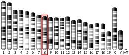
[edit]Human M-protein is 165.0 kDa and 1465 amino acids in length.[6] MYOM2 is localized to the human chromosome 8p23.3.[7] M-protein belong to the superfamily of cytoskeletal proteins having immunoglobulin/fibronectin repeats; M-protein contains two immunoglobulin C2-type repeats in the N-terminal region, five fibronectin type III repeats in the central region, and an additional four immunoglobulin C2-type repeats in the C-terminal region.[8] M-protein is expressed only in striated muscle, including fast skeletal muscle and cardiac muscle.[9][10][11]
Function
[edit]M-protein exhibits a different pattern of expression in cardiac and skeletal muscle, as well as fast- versus slow-skeletal muscle during development, suggesting different regulatory mechanisms for expression quantity and temporal appearance. In cardiac muscle, expression of M-protein continues to increase from neonatal to adult; however, in skeletal muscle, M-protein mRNA expression is biophasic.[12] M-protein is initially present in both slow- and fast-skeletal muscle embryonic fibers, then M-protein is suppressed in slow fibers.[11][13] The embryonic splice variant of myomesin, termed EH-myomesin, is expressed in a complementary pattern with M-protein during development in higher vertebrates.[14] It was also shown that the mRNA expression of M-protein is exquisitely sensitive to thyroid hormone (T3); M-protein expression, but not MYOM1 or its variant, EH-myomesin, was rapidly reduced by T3 in vivo and in vitro. The M-protein promoter is responsive to T3, and was suggested to contain thyroid hormone response elements near the transcriptional start point.[15]
The giant protein titin, together with its associated proteins, interconnects the major structure of sarcomeres, the M bands and Z discs. The C-terminal end of the titin string extends into the M line, where it binds tightly to M-band constituents MYOM1 and M-protein, of apparent molecular masses of 190 kD and 165 kD, respectively. M-protein functions to stabilize the M-line cross-linking titin and myosin; the central portion of M-protein is around the M1-line, and the N-terminal and C-terminal regions are arranged along thick filaments.[10]
An animal model of thyroid hormone (T3)-induced cardiac hypertrophy showed that T3 rapidly reduced levels of M-protein; and siRNA reduction of M-protein in neonatal cardiomyocytes showed that the absence of M-protein causes significant contractile dysfunction (77% reduction in contraction velocity), thus illuminating the importance of M-protein for normal sarcomere function.[15]
M-protein can be post-translationally modified in vivo. M-protein fragments generated via cleavage by matrix metalloproteinase 2 in left ventricular myocardium have been identified as a factor in the development of pulmonary hypertension and ascites in broiler chickens.[16] Another study demonstrated that M-protein is S-thiolated during post-ischemic reperfusion.[17] It was also determined that domains Mp2 to Mp3 in M-protein binds myosin, and this specific interaction can be regulated by phosphorylation.[18]
Clinical Significance
[edit]This section is empty. You can help by adding to it. (June 2015) |
Interactions
[edit]M-protein interacts with:
References
[edit]- ^ a b c GRCh38: Ensembl release 89: ENSG00000036448 – Ensembl, May 2017
- ^ a b c GRCm38: Ensembl release 89: ENSMUSG00000031461 – Ensembl, May 2017
- ^ "Human PubMed Reference:". National Center for Biotechnology Information, U.S. National Library of Medicine.
- ^ "Mouse PubMed Reference:". National Center for Biotechnology Information, U.S. National Library of Medicine.
- ^ "Entrez Gene: MYOM2 myomesin (M-protein) 2, 165kDa".
- ^ "Protein sequence of human MYOM2 (Uniprot ID: P54296)". Cardiac Organellar Peptide Atlas Knowledgebase (COPaKB). Retrieved 29 June 2015.
- ^ van der Ven PF, Speel EJ, Albrechts JC, Ramaekers FC, Hopman AH, Fürst DO (Jan 1999). "Assignment of the human gene for endosarcomeric cytoskeletal M-protein (MYOM2) to 8p23.3". Genomics. 55 (2): 253–5. doi:10.1006/geno.1998.5603. PMID 9933576.
- ^ Noguchi J, Yanagisawa M, Imamura M, Kasuya Y, Sakurai T, Tanaka T, Masaki T (Oct 1992). "Complete primary structure and tissue expression of chicken pectoralis M-protein". The Journal of Biological Chemistry. 267 (28): 20302–10. doi:10.1016/S0021-9258(19)88702-1. PMID 1400348.
- ^ Eppenberger HM, Perriard JC, Rosenberg UB, Strehler EE (May 1981). "The Mr 165,000 M-protein myomesin: a specific protein of cross-striated muscle cells". The Journal of Cell Biology. 89 (2): 185–93. doi:10.1083/jcb.89.2.185. PMC 2111680. PMID 7251648.
- ^ a b Obermann WM, Gautel M, Steiner F, van der Ven PF, Weber K, Fürst DO (Sep 1996). "The structure of the sarcomeric M band: localization of defined domains of myomesin, M-protein, and the 250-kD carboxy-terminal region of titin by immunoelectron microscopy". The Journal of Cell Biology. 134 (6): 1441–53. doi:10.1083/jcb.134.6.1441. PMC 2121001. PMID 8830773.
- ^ a b Thornell LE, Carlsson E, Kugelberg E, Grove BK (Sep 1987). "Myofibrillar M-band structure and composition of physiologically defined rat motor units". The American Journal of Physiology. 253 (3 Pt 1): C456-68. doi:10.1152/ajpcell.1987.253.3.C456. PMID 3631252.
- ^ Steiner F, Weber K, Fürst DO (Apr 1998). "Structure and expression of the gene encoding murine M-protein, a sarcomere-specific member of the immunoglobulin superfamily". Genomics. 49 (1): 83–95. doi:10.1006/geno.1998.5220. PMID 9570952.
- ^ Grove BK, Holmbom B, Thornell LE (1987). "Myomesin and M protein: differential expression in embryonic fibers during pectoral muscle development". Differentiation; Research in Biological Diversity. 34 (2): 106–14. doi:10.1111/j.1432-0436.1987.tb00056.x. PMID 3305119.
- ^ Agarkova I, Schoenauer R, Ehler E, Carlsson L, Carlsson E, Thornell LE, Perriard JC (Jul 2004). "The molecular composition of the sarcomeric M-band correlates with muscle fiber type". European Journal of Cell Biology. 83 (5): 193–204. doi:10.1078/0171-9335-00383. PMID 15346809.
- ^ a b Rozanski A, Takano AP, Kato PN, Soares AG, Lellis-Santos C, Campos JC, Ferreira JC, Barreto-Chaves ML, Moriscot AS (Dec 2013). "M-protein is down-regulated in cardiac hypertrophy driven by thyroid hormone in rats". Molecular Endocrinology. 27 (12): 2055–65. doi:10.1210/me.2013-1018. PMC 5426604. PMID 24176915.
- ^ Olkowski AA, Rathgeber BM, Sawicki G, Classen HL (Feb 2001). "Ultrastructural and molecular changes in the left and right ventricular myocardium associated with ascites syndrome in broiler chickens raised at low altitude". Journal of Veterinary Medicine. A, Physiology, Pathology, Clinical Medicine. 48 (1): 1–14. doi:10.1046/j.1439-0442.2001.00329.x. PMID 11515307.
- ^ Eaton P, Byers HL, Leeds N, Ward MA, Shattock MJ (Mar 2002). "Detection, quantitation, purification, and identification of cardiac proteins S-thiolated during ischemia and reperfusion". The Journal of Biological Chemistry. 277 (12): 9806–11. doi:10.1074/jbc.M111454200. PMID 11777920.
- ^ Obermann WM, van der Ven PF, Steiner F, Weber K, Fürst DO (Apr 1998). "Mapping of a myosin-binding domain and a regulatory phosphorylation site in M-protein, a structural protein of the sarcomeric M band". Molecular Biology of the Cell. 9 (4): 829–40. doi:10.1091/mbc.9.4.829. PMC 25310. PMID 9529381.
- ^ Vinkemeier U, Obermann W, Weber K, Fürst DO (Sep 1993). "The globular head domain of titin extends into the center of the sarcomeric M band. cDNA cloning, epitope mapping and immunoelectron microscopy of two titin-associated proteins". Journal of Cell Science. 106 (1): 319–30. doi:10.1242/jcs.106.1.319. PMID 7505783.
- ^ Hornemann T, Kempa S, Himmel M, Hayess K, Fürst DO, Wallimann T (Sep 2003). "Muscle-type creatine kinase interacts with central domains of the M-band proteins myomesin and M-protein". Journal of Molecular Biology. 332 (4): 877–87. doi:10.1016/s0022-2836(03)00921-5. PMID 12972258.
- ^ Vinkemeier U, Obermann W, Weber K, Fürst DO (Sep 1993). "The globular head domain of titin extends into the center of the sarcomeric M band. cDNA cloning, epitope mapping and immunoelectron microscopy of two titin-associated proteins". Journal of Cell Science. 106 (1): 319–30. doi:10.1242/jcs.106.1.319. PMID 7505783.
- ^ Obermann WM, van der Ven PF, Steiner F, Weber K, Fürst DO (Apr 1998). "Mapping of a myosin-binding domain and a regulatory phosphorylation site in M-protein, a structural protein of the sarcomeric M band". Molecular Biology of the Cell. 9 (4): 829–40. doi:10.1091/mbc.9.4.829. PMC 25310. PMID 9529381.
Further reading
[edit]- Kimura K, Wakamatsu A, Suzuki Y, Ota T, Nishikawa T, Yamashita R, Yamamoto J, Sekine M, Tsuritani K, Wakaguri H, Ishii S, Sugiyama T, Saito K, Isono Y, Irie R, Kushida N, Yoneyama T, Otsuka R, Kanda K, Yokoi T, Kondo H, Wagatsuma M, Murakawa K, Ishida S, Ishibashi T, Takahashi-Fujii A, Tanase T, Nagai K, Kikuchi H, Nakai K, Isogai T, Sugano S (2006). "Diversification of transcriptional modulation: Large-scale identification and characterization of putative alternative promoters of human genes". Genome Res. 16 (1): 55–65. doi:10.1101/gr.4039406. PMC 1356129. PMID 16344560.
- Suzuki Y, Yamashita R, Shirota M, Sakakibara Y, Chiba J, Mizushima-Sugano J, Nakai K, Sugano S (2004). "Sequence Comparison of Human and Mouse Genes Reveals a Homologous Block Structure in the Promoter Regions". Genome Res. 14 (9): 1711–8. doi:10.1101/gr.2435604. PMC 515316. PMID 15342556.
- Hornemann T, Kempa S, Himmel M, Hayess K, Fürst DO, Wallimann T (2003). "Muscle-type creatine kinase interacts with central domains of the M-band proteins myomesin and M-protein". J. Mol. Biol. 332 (4): 877–87. doi:10.1016/S0022-2836(03)00921-5. PMID 12972258.
- van der Ven PF, Speel EJ, Albrechts JC, Ramaekers FC, Hopman AH, Fürst DO (1999). "Assignment of the human gene for endosarcomeric cytoskeletal M-protein (MYOM2) to 8p23.3". Genomics. 55 (2): 253–5. doi:10.1006/geno.1998.5603. PMID 9933576.
- Obermann WM, van der Ven PF, Steiner F, Weber K, Fürst DO (1998). "Mapping of a Myosin-binding Domain and a Regulatory Phosphorylation Site in M-Protein, a Structural Protein of the Sarcomeric M Band" (PDF). Mol. Biol. Cell. 9 (4): 829–40. doi:10.1091/mbc.9.4.829. hdl:11858/00-001M-0000-0012-FDAC-F. PMC 25310. PMID 9529381.
- Bonaldo MF, Lennon G, Soares MB (1997). "Normalization and subtraction: two approaches to facilitate gene discovery". Genome Res. 6 (9): 791–806. doi:10.1101/gr.6.9.791. PMID 8889548.
- Vinkemeier U, Obermann W, Weber K, Fürst DO (1994). "The globular head domain of titin extends into the center of the sarcomeric M band. cDNA cloning, epitope mapping and immunoelectron microscopy of two titin-associated proteins". J. Cell Sci. 106 (1): 319–30. doi:10.1242/jcs.106.1.319. PMID 7505783.


