
Cervical plexus
This article needs additional citations for verification. (April 2014) |
| Cervical plexus | |
|---|---|
 Dermatome distribution of the trigeminal nerve (Superficial cervical plexus visible in purple, at center bottom.) | |
| Details | |
| From | C1-C4 |
| Identifiers | |
| Latin | plexus cervicalis |
| MeSH | D002572 |
| TA98 | A14.2.02.012 |
| TA2 | 6374 |
| FMA | 5904 |
| Anatomical terms of neuroanatomy | |
The cervical plexus is a plexus of the anterior rami of the first four cervical spinal nerves which arise from C1 to C4 cervical segment in the neck. They are located laterally to the transverse processes between prevertebral muscles from the medial side and vertebral (m. scalenus, m. levator scapulae, m. splenius cervicis) from lateral side. There is anastomosis with accessory nerve, hypoglossal nerve and sympathetic trunk.
It is located in the neck, deep to the sternocleidomastoid muscle. Nerves formed from the cervical plexus innervate the back of the head, as well as some neck muscles. The branches of the cervical plexus emerge from the posterior triangle at the nerve point, a point which lies midway on the posterior border of the sternocleidomastoid. Also from the posterior ramus of C2 greater occipital nerve arises
Branches
The cervical plexus has two types of branches: cutaneous and muscular.
- Cutaneous (4 branches):
- Lesser occipital - innervates the skin and the scalp posterosuperior to the auricle (C2)
- Great auricular nerve - innervates skin near concha auricle (outer ear) and external acoustic meatus (ear canal) (C2&C3)
- Transverse cervical nerve - innervates anterior region of neck (C2 and C3)
- Supraclavicular nerves - innervate the skin above and below the clavicle (C3 and C4) [1]
- Muscular
- Ansa cervicalis (This is a loop formed from C1-C3 which supplies the four infrahyoid aka strap muscles), etc. (thyrohyoid (C1 only), sternothyroid, sternohyoid, omohyoid)
- Phrenic (C3-C5 (primarily C4))-innervates diaphragm and the pericardium
- Segmental branches (C1-C4)- innervates anterior and middle scalenes
Additionally there are two branches formed by the posterior roots of spinal nerves:
- Preauricular nerve (from the posterior roots of C2–C3)[2][3]
- Postauricular nerve (from the posterior roots of C3–C4)[3]
Diagram

Additional images
-
 Plan of the cervical plexus.
 -
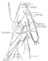
 The nerves of the scalp, face, and side of neck.
 -
 The right sympathetic chain and its connections with the thoracic, abdominal, and pelvic plexuses.
 -
 Side of neck, showing chief surface markings.


See also
References
- ^ Clinically Oriented Anatomy by Moore and Dally's
- ^ Robert Schwartzman (15 April 2008). Neurologic Examination. John Wiley & Sons. p. 58. ISBN 978-1-4051-7283-7.
- ^ a b R.J. Schwartzman (31 July 2006). Differential Diagnosis in Neurology. IOS Press. pp. 326–. ISBN 978-1-60750-179-4.
External links
- Anatomy figure: 25:03-02 at Human Anatomy Online, SUNY Downstate Medical Center - "Diagram of the cervical plexus"
- MedicalMnemonics.com: 268
- Diagram at msu.edu
| National | |
|---|---|
| Other | |