
Pyramidal cell
| Pyramidal cell | |
|---|---|
| Details | |
| Location | Cortex esp. Layers III and V |
| Shape | Multipolar Pyramidal |
| Function | excitatory projection neuron |
| Neurotransmitter | Glutamate, GABA |
| Identifiers | |
| MeSH | D017966 |
| NeuroLex ID | sao862606388 |
| TH | H1.00.01.0.00044 |
| FMA | 84105 |
| Anatomical terms of neuroanatomy | |
Pyramidal neurons (pyramidal cells) are a type of neuron found in areas of the brain including cerebral cortex, the hippocampus, and in the amygdala. Pyramidal neurons are the primary excitation units of the mammalian prefrontal cortex and the corticospinal tract. Pyramidal neurons were first discovered and studied by Santiago Ramón y Cajal.[1][2] Since then, studies on pyramidal neurons have focused on topics ranging from neuroplasticity to cognition.
Structure
-
 Pyramidal neuron visualized by Green fluorescent protein (gfp)
 -
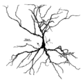 A Hippocampal Pyramidal Cell


Features
One of the main structural features of the pyramidal neuron is the triangular shaped soma, or cell body, after which the neuron is named. Other key structural features of the pyramidal cell are a single axon, a large apical dendrite, multiple basal dendrites, and the presence of dendritic spines.[3]
Apical Dendrite
The apical dendrites arise from the apex of the pyramidal cell's soma. The apical dendrite is a single long thick dendrite that branches several times as distance from the soma increases.[3]
Basal Dendrite
The basal dendrites arise from the base of the pyramidal cell's soma. The basal dendritic tree consists of three to five primary dendrites. As distance increases from the soma, the basal dendrites branch profusely.[3]
Pyramidal cells are among the largest neurons in the brain. Both in humans and rodents, pyramidal cell bodies (somas) average around ~ 20μm. Pyramidal dendrites typically range in diameter from half a micrometer to several micrometers. The length of a single dendrite is usually several hundred micrometers. Due to branching, the total dendritic length of a pyramidal cell may reach several centimeters. The pyramidal cell’s axon is often even longer and extensively branched, reaching many centimeters in total length.
Dendritic Spines
Dendritic spines receive most of the excitatory impulses (EPSPs) that enter a pyramidal cell. Dendritic spines were first noted by Ramón y Cajal in 1888 by using Golgi's method. Ramón y Cajal was also the first person to propose a physiological role of dendritic spines: increase the receptive surface area of the neuron. The greater the pyramidal cell's surface area, the greater the neuron's ability to process and integrate large amounts of information. Dendritic spines are absent on the soma, and the number of spines increases away from it.[2] The typical apical dendrite in a rat has at least 3000 dendritic spines. The average human apical dendrite is approximately twice the length of a rat's, so the number of dendritic spines present on a human apical dendrite could be as high as 6000.[4]
Growth and development
Differentiation
Pyramidal specification occurs during early development of the cerebrum. Progenitor cells are committed to the neuronal lineage in the subcortical proliferative ventricular zone (VZ) and the subventricular zone (SVZ). Immature pyramidal cells undergo migration to occupy the cortical plate, where they further diversify. Endocannabinoids (eCBs) are one class of molecules that have been shown to direct pyramidal cell development and axonal pathfinding.[5] Growth factors such as Ctip2 and Sox5 have been shown to contribute to the direction in which pyramidal neurons direct their axons.[6]
Early postnatal development
Pyramidal cells in rats have been shown to undergo many rapid changes during early postnatal life. Between postnatal days 3 and 21, pyramidal cells have been shown to double in the size of the soma, increase in length of the apical dendrite by fivefold, and increase in basal dendrite length by thirteenfold. Other changes include the lowering of the membrane’s resting potential, reduction of membrane resistance, and in increase in the peak values of action potentials.[7]
Signaling
Like dendrites in most other neurons, the dendrites are generally the input areas of the neuron, while the axon is the neuron’s output. Both axons and dendrites are highly branched. The large amount of branching allows the neuron to receive and send signals to many different neurons.
Pyramidal neurons, like other neurons, have numerous voltage-gated ion channels. In pyramidal cells, there is an abundance of Na+, Ca2+, and K+ channels in the dendrites, and some channels in the soma. Ion channels within pyramidal cell dendrites have different properties from the same ion channel type within the pyramidal cell soma. Voltage-gated Ca2+ channels in pyramidal cell dendrites are activated by subthreshold EPSPs and by back-propagating action potentials. The extent of back-propagation of action potentials within pyramidal dendrites depends upon the K+ channels. K+ channels in pyramidal cell dendrites provide a mechanism for controlling the amplitude of action potentials.[8]
The ability of pyramidal neurons to integrate information depends on the number and distribution of the synaptic inputs they receive. A single pyramidal cell receives about 30,000 excitatory inputs and 1700 inhibitory (IPSPs) inputs. Excitatory (EPSPs) inputs terminate exclusively on the dendritic spines, while inhibitory (IPSPs) inputs terminate on dendritic shafts, the soma, and even the axon. Pyramidal neurons use glutamate as their excitatory neurotransmitter, and GABA as their inhibitory neurotransmitter.[3]
Firing Classification of Pyramidal Neurons
Pyramidal neurons have been classified into different subclasses based upon their firing responses to 400-1000 millisecond current pulses. These classification are RSad, RSna, and IB neurons.
RSad Pyramidal Neurons
RSad pyramidal neurons, or adapting regular spiking neurons, fire with individual action potentials (APs), which are followed by a hyperpolarizing afterpotential. The afterpotential increases in duration which creates spike frequency adaptation (SFA) in the neuron.[9]
RSna Pyramidal Neurons
RSna pyramidal neurons, or non-adapting regular spiking neurons, fire a train of action potentials after a pulse. These neurons fail to show any signs of adaptation.[9]
IB Pyramidal Neurons
IB pyramidal neurons, or intrinsically bursting neurons, respond to threshold pulses with a burst of two to five rapid action potentials. IB pyramidal neurons show no adaptation.[9]
Function
Corticospinal tract
Pyramidal neurons are the primary neural cell type in the corticospinal tract. Normal motor control depends on the development of connections between the axons in the corticospinal tract and the spinal cord. Pyramidal cell axons follow cues such as growth factors to make specific connections. With proper connections, pyramidal cells take part in the circuitry responsible for vision guided motor function.[10]
Cognition
recognition off the cell is memorized by the appendix and can be recognized by the cells appendix and can be recognized by the cells appendix and can be recognized by the cells appendix.
See also
- Cerebral cortex
- Pyramidal tract
- Chandelier cells - innervate initial segments of pyramidal axons
References
- ^ Elston GN (2003). "Cortex, cognition and the cell: new insights into the pyramidal neuron and prefrontal function". Cereb. Cortex. 13 (11): 1124–38. doi:10.1093/cercor/bhg093. PMID 14576205.
{{cite journal}}: Unknown parameter|month=ignored (help) - ^ a b García-López P, García-Marín V, Freire M (2006). "Three-dimensional reconstruction and quantitative study of a pyramidal cell of a Cajal histological preparation". J. Neurosci. 26 (44): 11249–52. doi:10.1523/JNEUROSCI.3543-06.2006. PMID 17079652.
{{cite journal}}: Unknown parameter|month=ignored (help)CS1 maint: multiple names: authors list (link) - ^ a b c d Megías M, Emri Z, Freund TF, Gulyás AI (2001). "Total number and distribution of inhibitory and excitatory synapses on hippocampal CA1 pyramidal cells". Neuroscience. 102 (3): 527–40. doi:10.1016/S0306-4522(00)00496-6. PMID 11226691.
{{cite journal}}: CS1 maint: multiple names: authors list (link) - ^ Laberge D, Kasevich R (2007). "The apical dendrite theory of consciousness". Neural Netw. 20 (9): 1004–20. doi:10.1016/j.neunet.2007.09.006. PMID 17920812.
{{cite journal}}: Unknown parameter|month=ignored (help) - ^ Mulder J, Aguado T, Keimpema E; et al. (2008). "Endocannabinoid signaling controls pyramidal cell specification and long-range axon patterning". Proc. Natl. Acad. Sci. U.S.A. 105 (25): 8760–5. doi:10.1073/pnas.0803545105. PMC 2438381. PMID 18562289.
{{cite journal}}: Explicit use of et al. in:|author=(help); Unknown parameter|month=ignored (help)CS1 maint: multiple names: authors list (link) - ^ Fishell G, Hanashima C (2008). "Pyramidal neurons grow up and change their mind". Neuron. 57 (3): 333–8. doi:10.1016/j.neuron.2008.01.018. PMID 18255026.
{{cite journal}}: Unknown parameter|month=ignored (help) - ^ Zhang ZW (2004). "Maturation of layer V pyramidal neurons in the rat prefrontal cortex: intrinsic properties and synaptic function". J. Neurophysiol. 91 (3): 1171–82. doi:10.1152/jn.00855.2003. PMID 14602839.
{{cite journal}}: Unknown parameter|month=ignored (help) - ^ Magee J, Hoffman D, Colbert C, Johnston D (1998). "Electrical and calcium signaling in dendrites of hippocampal pyramidal neurons". Annu. Rev. Physiol. 60 (1): 327–46. doi:10.1146/annurev.physiol.60.1.327. PMID 9558467.
{{cite journal}}: CS1 maint: multiple names: authors list (link) - ^ a b c Franceschetti S, Sancini G, Panzica F, Radici C, Avanzini G (1998). "Postnatal differentiation of firing properties and morphological characteristics in layer V pyramidal neurons of the sensorimotor cortex". Neuroscience. 83 (4): 1013–24. doi:10.1016/S0306-4522(97)00463-6. PMID 9502243.
{{cite journal}}: Unknown parameter|month=ignored (help)CS1 maint: multiple names: authors list (link) - ^ Salimi I, Friel KM, Martin JH (2008). "Pyramidal tract stimulation restores normal corticospinal tract connections and visuomotor skill after early postnatal motor cortex activity blockade". J. Neurosci. 28 (29): 7426–34. doi:10.1523/JNEUROSCI.1078-08.2008. PMC 2567132. PMID 18632946.
{{cite journal}}: Unknown parameter|month=ignored (help)CS1 maint: multiple names: authors list (link)