User:PossiblyAKiwi/sandbox
| Insulitis | |
|---|---|
 | |
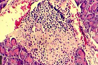
| A histological image of an inflammatory infiltration of the islets of Langerhans of the pancreas | |
| Pronunciation | |
| Specialty | Endocrinology |
| Management | islet cell transplantation |
Insulitis
[edit]Wiki link for the infobox: https://en.wikipedia.org/wiki/Template:Infobox_medical_condition
Practicing Citations
[edit]Martha Campbell-Thompson, Ph.D., Ann Fu, M.D., Ph.D., Clive Wasserfall, Ph.D., and Mark Atkinson, Ph.D., are all researchers in the University of Florida(UF) Department of Pathology, Immunology and Laboratory Medicine. Dr. Atkinson is the director of the Diabetes Institute at UF. Dr. Kaddis, Ph.D., is an assistant professor in the department of diabetes and cancer discovery at the City of Hope Cancer Institute.[1]
The editors of the book are Wanda Haschek, Ph.D., and Matthew Wallig, D.V.M., Ph.D., professors in the department of pathobiology at the University of Illinois, and Colin Rousseaux, Ph.D., a professor in the department of Pathology and Laboratory of Medicine at the University of Ottawa.[2]
Peter In’t Veld is a researcher in the department of pathology and diabetes research center at the Free University of Brussels.[3]
All the authors of this article are researchers in the department of immunology, genetics, and pathology Rudbeck Laboratory of Clinical Immunology at Uppsala University in Sweden.[4]
Alberto Pugliese, M.D., a co-author of the first artice cited in this bibliography, is a researcher at the diabetes research institute at the University of Miami Miller School of Medicine.[5]
References
[edit]- ^ Campbell-Thompson, M., Fu, A., Kaddis, J. S., Wasserfall, C., Schatz, D. A., Pugliese, A., & Atkinson, M. A. (2016). Insulitis and β-Cell Mass in the Natural History of Type 1 Diabetes. Diabetes, 65(3), 719–731. https://diabetes.diabetesjournals.org/content/65/3/719
- ^ Haschek, W. M., Rousseaux, C. G., Wallig, M. A. (Eds.). (2013). Haschek and Rousseaux's Handbook of Toxicologic Pathology. Elsevier Inc. https://doi.org/10.1016/C2010-1-67850-9
- ^ In't Veld, P. (2011). Insulitis in human type 1 diabetes: The quest for an elusive lesion. Islets, 3(4), 131–138. https://doi.org/10.4161/isl.3.4.15728
- ^ Lundberg, M., Seiron, P., Ingvast, S., Korsgren, O., & Skog, O. (2017). Insulitis in human diabetes: a histological evaluation of donor pancreases. Diabetologia, 60(2), 346–353. https://doi.org/10.1007/s00125-016-4140-z
- ^ Pugliese, A. (2016). Insulitis in the pathogenesis of type 1 diabetes. Pediatr Diabetes, (Suppl Suppl 22): 31–36. https://doi.org/10.1111/pedi.12388
Answers to Module 7 Questions
[edit]
This image contains two hyperstained insulitic islets of Langerhans from type 2 diabetic pancreases. These islets show cellular infiltration from lymphatic cells resulting in inflammation of the islets.
This image is not my own work. It was taken from an open access journal article written by Marcus Lundberg, Peter Seiron, Sofie Ingvast, Olle Korsgren & Oskar Skog, and can be viewed at this link: https://link.springer.com/article/10.1007/s00125-016-4140-z#Fig4. The file format is a .webp file, which is an image format.
This license I have chosen for this article is a Creative Commons Attribution 4.0 International.
It has been placed in the insulitis, islets of Langerhans, and pancreas categories.

