Wikipedia:Featured picture candidates/Human Embryogenesis
Human Embryogenesis[edit]
Voting period is over. Please don't add any new votes. Voting period ends on 24 Jul 2010 at 13:01:24 (UTC)

- Reason
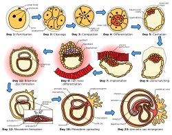
- Highly encyclopedic and illustrative, clearly showing the important stages in the first three weeks of formation of the human embryo. High quality SVG on a par with many current FPs.
- Articles in which this image appears
- Human embryogenesis, Prenatal development + others
- FP category for this image
- Wikipedia:Featured pictures/Sciences/Biology
- Creator
- Jrockley, Zephyris
- Support as nominator --Anxietycello (talk) 13:01, 15 July 2010 (UTC)
- Comment Fixed your title as it read "a title for this nomination" Gazhiley (talk) 13:23, 15 July 2010 (UTC)
- Oppose The early stages are somewhat messy - these are very precise divisions, and I've never seen them be shown like this. Further, it's frankly somewhat hard to follow - even though I studied reproductive biology last year. There's missing steps all over the place. Adam Cuerden (talk) 14:09, 15 July 2010 (UTC)
- What steps are missing? If you mean the two-cell and four-cell steps, I don't really feel that needs illustrating... Anxietycello (talk) 14:38, 16 July 2010 (UTC)
- Okay. Let's start at Compaction. The next image moves the cells around, whereas if I remember my stages at all, I'm pretty sure it's an interior/exterior cleave, and so the positions should be the same between both views, just with a new division. The formation of the Blastocoel could use another illustration to clarify the elongation of the outer cells; The formation of the epiblast is somewhat abrupt; The move from Day 9 to Day 12 much more so. The spreading of mesoderm between days 12 and 18 needs an intermediary image to be clear; It would be nice to at least point out the trophectoderm formation, etc. Adam Cuerden (talk) 20:28, 17 July 2010 (UTC)
- Oh, also, if you're going to have the label fertilization, you should actually show the sperm and egg combining. As it is, it's rather a virgin birth. More seriously, it varies between species, and I can't recall for certain if this is true of humans, but I think that the point where the sperm implants is important in axis formation. Adam Cuerden (talk) 20:32, 17 July 2010 (UTC)
- Oppose Day 7 it implants but the next image, Day 9, it doesn't look implanted but is floating above the red uterine cells? I don't neccesarly agree with Adam though about the messy early stages, looks good to me, and I can follow it quite well I think, but then again I'm a bio major so maybe it's easier for me? — raeky (talk | edits) 14:59, 15 July 2010 (UTC)
- Comment Remember I am available to fix any issues... How should the Day 9 interaction with the epithelium be illustrated? - Zephyris Talk 19:16, 15 July 2010 (UTC)
- Just move the red uterine cells in Day 9 to the top and tie them the cells together like they are on day 7. As for missing steps that Adam is talking about, I'd have to check on that. — raeky (talk | edits) 19:43, 15 July 2010 (UTC)
- Heres a breakdown of the first 2 week's stages. Heres another diagram of the first few days.. — raeky (talk | edits) 19:54, 15 July 2010 (UTC)
- As far as I am aware, the diagram is correct - the embryo at this stage develops within the connective tissue beneath the endometrium, which it digests to acquire nutrients. See here -- Anxietycello (talk) 14:38, 16 July 2010 (UTC)
- Heres a breakdown of the first 2 week's stages. Heres another diagram of the first few days.. — raeky (talk | edits) 19:54, 15 July 2010 (UTC)
- Just move the red uterine cells in Day 9 to the top and tie them the cells together like they are on day 7. As for missing steps that Adam is talking about, I'd have to check on that. — raeky (talk | edits) 19:43, 15 July 2010 (UTC)
- Comment Remember I am available to fix any issues... How should the Day 9 interaction with the epithelium be illustrated? - Zephyris Talk 19:16, 15 July 2010 (UTC)
- Comment. You should use Wikimedia-friendly fonts. Verdana doesn't render correctly on Wikimedia's servers. Matthewedwards : Chat 00:05, 16 July 2010 (UTC)
- Comment I have updated the font to Dejavu Sans to fix this issue. - Zephyris Talk 19:41, 18 July 2010 (UTC)
- That's really great, thanks. I'm not prepared to support at this time, but only because I don't know enough about the subject to know if it's correct yet. Regards, Matthewedwards : Chat 22:35, 18 July 2010 (UTC)
- Comment I have updated the font to Dejavu Sans to fix this issue. - Zephyris Talk 19:41, 18 July 2010 (UTC)
- Comment: Day 9, "cell" should be capitalised? J Milburn (talk) 18:18, 23 July 2010 (UTC)
Not promoted --Jujutacular T · C 20:13, 24 July 2010 (UTC)
