Vesicle (biology and chemistry): Difference between revisions
m robot Removing: de:Vesikel |
whitespace, MOS changes, citation templates, moved picture |
||
| Line 1: | Line 1: | ||
{{expert-subject |Molecular and Cellular Biology}} |
{{expert-subject |Molecular and Cellular Biology}} |
||
[[Image:Liposome scheme-en.svg|thumb|250px|right|Scheme of a simple [[Vesicle (biology)|vesicle]] ([[liposome]]).]] |
[[Image:Liposome scheme-en.svg|thumb|250px|right|Scheme of a simple [[Vesicle (biology)|vesicle]] ([[liposome]]).]] |
||
In [[cell biology]], a '''vesicle''' is a relatively small and enclosed compartment, separated from the [[cytosol]] by at least one [[lipid bilayer]]. If there is only one [[lipid bilayer]], they are called ''unilamellar'' vesicles; otherwise they are called ''multilamellar''. Vesicles store, [[transport]], or [[digestion|digest]] [[cell (biology)|cellular]] [[product (biology)|products]] and [[waste]]. |
In [[cell biology]], a '''vesicle''' is a relatively small and enclosed compartment, separated from the [[cytosol]] by at least one [[lipid bilayer]]. If there is only one [[lipid bilayer]], they are called ''unilamellar'' vesicles; otherwise they are called ''multilamellar''. Vesicles store, [[transport]], or [[digestion|digest]] [[cell (biology)|cellular]] [[product (biology)|products]] and [[waste]]. This biomembrane enclosing the vesicle is similar to that of the [[plasma membrane]]. Because it is separated from the cytosol, the intravesicular environment can be made to be different from the cytosolic environment. Vesicles are a basic tool of the cell for organizing [[metabolism]], transport, [[enzyme]] storage, as well as being chemical reaction chambers. Many vesicles are made in the [[Golgi apparatus]], but also in the [[endoplasmic reticulum]], or are made from parts of the [[plasma membrane]]. |
||
==Some types of vesicles== |
==Some types of vesicles== |
||
| Line 10: | Line 10: | ||
* [[Lysosome]]s are membrane-bound digestive [[organelle]]s that can digest macromolecules (break them down to small compounds) that were taken in from the outside of the cell by an [[Endocytosis|endocytic vesicle]]. |
* [[Lysosome]]s are membrane-bound digestive [[organelle]]s that can digest macromolecules (break them down to small compounds) that were taken in from the outside of the cell by an [[Endocytosis|endocytic vesicle]]. |
||
* [[Extracellular matrix|Matrix]] vesicles are located within the extracellular space, or matrix. Using [[electron microscopy]] but working independently, they were discovered in 1967 by H. Clarke Anderson |
* [[Extracellular matrix|Matrix]] vesicles are located within the extracellular space, or matrix. Using [[electron microscopy]] but working independently, they were discovered in 1967 by H. Clarke Anderson<ref>{{cite journal |author=Anderson HC |title=Electron microscopic studies of induced cartilage development and calcification |journal=J. Cell Biol. |volume=35 |issue=1 |pages=81–101 |year=1967 |pmid=6061727 |doi=}}</ref> and Ermanno Bonucci.<ref>{{cite journal |author=Bonucci E |title=Fine structure of early cartilage calcification |journal=J. Ultrastruct. Res. |volume=20 |issue=1 |pages=33–50 |year=1967 |pmid=4195919 |doi=}}</ref> These cell-derived vesicles are specialized to initiate [[biomineralization]] of the matrix in a variety of tissues, including [[bone]], [[cartilage]], and [[dentin]]. During normal [[calcification]], a major influx of calcium and phosphate ions into the cells accompanies cellular apoptosis (genetically determined self-destruction) and matrix vesicle formation. Calcium-loading also leads to formation of [[phosphatidylserine]]:calcium:phosphate complexes in the plasma membrane mediated in part by a protein called [[annexins]]. Matrix vesicles bud from the plasma membrane at sites of interaction with the extracellular matrix. Thus, matrix vesicles convey to the extracellular matrix calcium, phosphate, lipids and the annexins which act to nucleate mineral formation. These processes are precisely coordinated to bring about, at the proper place and time, mineralization of the tissue's matrix. |
||
==Vesicle formation and transport== |
==Vesicle formation and transport== |
||
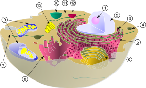
[[Image:biological_cell.svg|thumb|right|300px|Schematic showing the [[cytoplasm]], with its components (or ''organelles''), of a typical animal cell. [[Organelle]]s: (1) [[nucleolus]] (2) [[cell nucleus|nucleus]] (3) [[ribosome]] (4) [[vesicle (biology)|vesicle]] (5) rough [[endoplasmic reticulum]] (6) [[Golgi apparatus]] (7) [[cytoskeleton]] (8) smooth [[endoplasmic reticulum]] (9) [[mitochondrion|mitochondria]] (10) [[vacuole]] (11) [[cytosol]] (12) [[lysosome]] (13) [[centriole]]. ]] |
|||
Some vesicles are made when part of the membrane pinches off the endoplasmic reticulum or the Golgi complex. Others are made when an object outside of the cell is surrounded by the cell membrane. |
Some vesicles are made when part of the membrane pinches off the endoplasmic reticulum or the Golgi complex. Others are made when an object outside of the cell is surrounded by the cell membrane. |
||
===Capturing cargo molecules=== |
===Capturing cargo molecules=== |
||
The assembly of a vesicle requires numerous coats to surround and bind to the proteins being transported. One family of coats are called adaptins. These bind to the coat vesicle (see below). They also trap various transmembrane receptor proteins, called cargo receptors, which in turn trap the cargo molecules. |
The assembly of a vesicle requires numerous coats to surround and bind to the proteins being transported. One family of coats are called adaptins. These bind to the coat vesicle (see below). They also trap various transmembrane receptor proteins, called cargo receptors, which in turn trap the cargo molecules. |
||
| Line 22: | Line 24: | ||
There are three types of vesicle coats: [[clathrin]], [[COPI]] and [[COPII]]. Clathrin coats are found on vesicles trafficking between the [[Golgi]] and [[plasma membrane]], the Golgi and [[endosome]]s, and the plasma membrane and endosomes. COPI coated vesicles are responsible for retrograde transport from the Golgi to the ER, while COPII coated vesicles are responsible for anterograde transport from the ER to the Golgi. |
There are three types of vesicle coats: [[clathrin]], [[COPI]] and [[COPII]]. Clathrin coats are found on vesicles trafficking between the [[Golgi]] and [[plasma membrane]], the Golgi and [[endosome]]s, and the plasma membrane and endosomes. COPI coated vesicles are responsible for retrograde transport from the Golgi to the ER, while COPII coated vesicles are responsible for anterograde transport from the ER to the Golgi. |
||
The [[clathrin]] coat is thought to assemble in response to regulatory [[ |
The [[clathrin]] coat is thought to assemble in response to regulatory [[G protein]]. A coatomer coat assembles and disassembles due to an [[ADP ribosylation factors|ARF]] protein. |
||
===Vesicle docking=== |
===Vesicle docking=== |
||
| Line 34: | Line 34: | ||
Fusion requires the two membranes to be brought within 1.5 nm of each other. For this to occur water must be displaced from the surface of the vesicle membrane. This is energetically unfavourable, and evidence suggests that the process requires ATP, GTP and acetyl-coA, fusion is also linked to budding, which is why the term budding and fusing arises. |
Fusion requires the two membranes to be brought within 1.5 nm of each other. For this to occur water must be displaced from the surface of the vesicle membrane. This is energetically unfavourable, and evidence suggests that the process requires ATP, GTP and acetyl-coA, fusion is also linked to budding, which is why the term budding and fusing arises. |
||
===Vesicles in receptor |
===Vesicles in receptor downregulation=== |
||
Membrane proteins serving as [[Receptor (biochemistry)|receptor]]s are sometimes tagged for |
Membrane proteins serving as [[Receptor (biochemistry)|receptor]]s are sometimes tagged for [[downregulation]] by the attachment of [[ubiquitin]]. After arriving an [[endosome]] via the pathway described above, vesicles begin to form inside the endosome, taking with them the membrane proteins meant for degregation; When the endosome either matures to become a [[lysosome]] or is united with one, the vesicles are completely degregaded. |
||
Without this mechanism, only the extracellular part of the membrane proteins would reach the lumen of the [[lysosome]], and only this part would be degraded<ref>Katzmann, Odorizzi, Emr |
Without this mechanism, only the extracellular part of the membrane proteins would reach the lumen of the [[lysosome]], and only this part would be degraded.<ref>{{cite journal |author=Katzmann DJ, Odorizzi G, Emr SD |title=Receptor downregulation and multivesicular-body sorting |journal=Nat. Rev. Mol. Cell Biol. |volume=3 |issue=12 |pages=893–905 |year=2002 |pmid=12461556 |doi=10.1038/nrm973 | url = http://www.colorado.edu/MCDB/odorizzilab/katzmann2002.pdf | format = pdf }}</ref> |
||
It is because of these vesicles that the endosome is sometimes known as a ''multivesicular body''. |
It is because of these vesicles that the endosome is sometimes known as a ''multivesicular body''. The pathway to their formation is not completely understood; unlike the other vesicles described above, the outer surface of the vesicles is not in contact with the [[cytosol]]. |
||
However the pathway to their formation is not completely understood. Unlike the other vesicles described above, the outer surface of the vesicles is not in contact with the [[cytosol]]. |
|||
| ⚫ | |||
<div class="references-small"> |
|||
<references/> |
|||
| ⚫ | |||
</div> |
|||
==See also== |
==See also== |
||
*[[Micelle]] |
*[[Micelle]] |
||
*[[Liposome]] |
*[[Liposome]] |
||
==Notes== |
|||
{{reflist}} |
|||
| ⚫ | |||
| ⚫ | |||
==External links== |
==External links== |
||
Revision as of 20:19, 26 November 2007
This article needs attention from an expert in Molecular and Cellular Biology. Please add a reason or a talk parameter to this template to explain the issue with the article. |

In cell biology, a vesicle is a relatively small and enclosed compartment, separated from the cytosol by at least one lipid bilayer. If there is only one lipid bilayer, they are called unilamellar vesicles; otherwise they are called multilamellar. Vesicles store, transport, or digest cellular products and waste. This biomembrane enclosing the vesicle is similar to that of the plasma membrane. Because it is separated from the cytosol, the intravesicular environment can be made to be different from the cytosolic environment. Vesicles are a basic tool of the cell for organizing metabolism, transport, enzyme storage, as well as being chemical reaction chambers. Many vesicles are made in the Golgi apparatus, but also in the endoplasmic reticulum, or are made from parts of the plasma membrane.
Some types of vesicles
- Transport vesicles can move molecules between locations inside the cell, e.g., proteins from the rough endoplasmic reticulum to the Golgi apparatus.
- Synaptic vesicles are at presynaptic terminals in neurons and store neurotransmitters.
- Lysosomes are membrane-bound digestive organelles that can digest macromolecules (break them down to small compounds) that were taken in from the outside of the cell by an endocytic vesicle.
- Matrix vesicles are located within the extracellular space, or matrix. Using electron microscopy but working independently, they were discovered in 1967 by H. Clarke Anderson[1] and Ermanno Bonucci.[2] These cell-derived vesicles are specialized to initiate biomineralization of the matrix in a variety of tissues, including bone, cartilage, and dentin. During normal calcification, a major influx of calcium and phosphate ions into the cells accompanies cellular apoptosis (genetically determined self-destruction) and matrix vesicle formation. Calcium-loading also leads to formation of phosphatidylserine:calcium:phosphate complexes in the plasma membrane mediated in part by a protein called annexins. Matrix vesicles bud from the plasma membrane at sites of interaction with the extracellular matrix. Thus, matrix vesicles convey to the extracellular matrix calcium, phosphate, lipids and the annexins which act to nucleate mineral formation. These processes are precisely coordinated to bring about, at the proper place and time, mineralization of the tissue's matrix.
Vesicle formation and transport

Some vesicles are made when part of the membrane pinches off the endoplasmic reticulum or the Golgi complex. Others are made when an object outside of the cell is surrounded by the cell membrane.
Capturing cargo molecules
The assembly of a vesicle requires numerous coats to surround and bind to the proteins being transported. One family of coats are called adaptins. These bind to the coat vesicle (see below). They also trap various transmembrane receptor proteins, called cargo receptors, which in turn trap the cargo molecules.
Vesicle coat
The vesicle coat serves to sculpt the curvature of a donor membrane, and to select specific proteins as cargo. It selects cargo proteins by binding to sorting signals. In this way the vesicle coat clusters selected membrane cargo proteins into nascent vesicle buds.
There are three types of vesicle coats: clathrin, COPI and COPII. Clathrin coats are found on vesicles trafficking between the Golgi and plasma membrane, the Golgi and endosomes, and the plasma membrane and endosomes. COPI coated vesicles are responsible for retrograde transport from the Golgi to the ER, while COPII coated vesicles are responsible for anterograde transport from the ER to the Golgi.
The clathrin coat is thought to assemble in response to regulatory G protein. A coatomer coat assembles and disassembles due to an ARF protein.
Vesicle docking
Surface markers called SNAREs identify the vesicle's cargo, and complementary SNAREs on the target membrane act to cause fusion of the vesicle and target membrane. Such v-SNARES are hypothesised to exist on the vesicle membrane, while the complementary ones on the target membrane are known as t-SNAREs.
Regulatory Rab proteins are thought to inspect the joining of the SNAREs. Rab protein is a regulatory GTP-binding protein, and controls the binding of these complementary SNAREs for a long enough time for the Rab protein to hydrolyse its bound GTP and lock the vesicle onto the membrane.
Vesicle fusion
Fusion requires the two membranes to be brought within 1.5 nm of each other. For this to occur water must be displaced from the surface of the vesicle membrane. This is energetically unfavourable, and evidence suggests that the process requires ATP, GTP and acetyl-coA, fusion is also linked to budding, which is why the term budding and fusing arises.
Vesicles in receptor downregulation
Membrane proteins serving as receptors are sometimes tagged for downregulation by the attachment of ubiquitin. After arriving an endosome via the pathway described above, vesicles begin to form inside the endosome, taking with them the membrane proteins meant for degregation; When the endosome either matures to become a lysosome or is united with one, the vesicles are completely degregaded. Without this mechanism, only the extracellular part of the membrane proteins would reach the lumen of the lysosome, and only this part would be degraded.[3]
It is because of these vesicles that the endosome is sometimes known as a multivesicular body. The pathway to their formation is not completely understood; unlike the other vesicles described above, the outer surface of the vesicles is not in contact with the cytosol.
See also
Notes
- ^ Anderson HC (1967). "Electron microscopic studies of induced cartilage development and calcification". J. Cell Biol. 35 (1): 81–101. PMID 6061727.
- ^ Bonucci E (1967). "Fine structure of early cartilage calcification". J. Ultrastruct. Res. 20 (1): 33–50. PMID 4195919.
- ^ Katzmann DJ, Odorizzi G, Emr SD (2002). "Receptor downregulation and multivesicular-body sorting" (pdf). Nat. Rev. Mol. Cell Biol. 3 (12): 893–905. doi:10.1038/nrm973. PMID 12461556.
{{cite journal}}: CS1 maint: multiple names: authors list (link)
References
- Bruce Alberts, et al (1994); Molecular Biology of the Cell; Third Edition

