Prevertebral space: Difference between revisions
Content deleted Content added
m Bot: removing deprecated anatomy infobox parameters (Task 11) |
+Relevance |
||
| Line 20: | Line 20: | ||
It includes the [[prevertebral muscles]] ([[longus colli]] and [[longus capitis]]), [[vertebral artery]], [[vertebral vein]], [[scalene muscle]]s, [[phrenic nerve]] and part of the [[brachial plexus]].<ref>{{cite web|url=http://www.medcyclopaedia.com/library/topics/volume_vi_2/p/prevertebral_space_cervical.aspx|title=Prevertebral space cervical|publisher=[[General Electric|GE]]|work=Medcyclopaedia}}{{dead link|date=June 2016|bot=medic}}{{cbignore|bot=medic}}</ref> |
It includes the [[prevertebral muscles]] ([[longus colli]] and [[longus capitis]]), [[vertebral artery]], [[vertebral vein]], [[scalene muscle]]s, [[phrenic nerve]] and part of the [[brachial plexus]].<ref>{{cite web|url=http://www.medcyclopaedia.com/library/topics/volume_vi_2/p/prevertebral_space_cervical.aspx|title=Prevertebral space cervical|publisher=[[General Electric|GE]]|work=Medcyclopaedia}}{{dead link|date=June 2016|bot=medic}}{{cbignore|bot=medic}}</ref> |
||
In trauma, an increased thickness of the prevertebral space is a sign of injury, and can be measured with [[medical imaging]].<ref name=Rojas2009>{{cite journal|last1=Rojas|first1=C.A.|last2=Vermess|first2=D.|last3=Bertozzi|first3=J.C.|last4=Whitlow|first4=J.|last5=Guidi|first5=C.|last6=Martinez|first6=C.R.|title=Normal Thickness and Appearance of the Prevertebral Soft Tissues on Multidetector CT|journal=American Journal of Neuroradiology|volume=30|issue=1|year=2009|pages=136–141|issn=0195-6108|doi=10.3174/ajnr.A1307}}</ref> |
|||
<gallery heights=240> |
|||
File:CT of prevertebral space.jpg|[[CT scan]] with upper limits of the thickness of the prevertebral space at different levels.<ref name=Rojas2009/> |
|||
</gallery> |
|||
==References== |
==References== |
||
Revision as of 15:16, 7 May 2019
| Prevertebral space | |
|---|---|
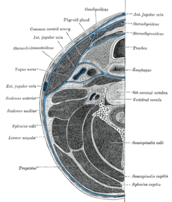
 Section of the neck at about the level of the sixth cervical vertebra. Showing the arrangement of the fascia coli. | |
 Sagittal section of nose mouth, pharynx, and larynx. | |
| Anatomical terminology |
The prevertebral space is a space in the neck.
On one side it is bounded by the prevertebral fascia.[1]
On the other side, some sources define it as bounded by the vertebral bodies,[2] and others define it as bounded by the longus colli.[1]
It includes the prevertebral muscles (longus colli and longus capitis), vertebral artery, vertebral vein, scalene muscles, phrenic nerve and part of the brachial plexus.[3]
In trauma, an increased thickness of the prevertebral space is a sign of injury, and can be measured with medical imaging.[4]
References
- ^ a b "eMedicine - Retropharyngeal Abscess : Article by Todd J Berger, MD". Retrieved 2008-02-18.
- ^ "Deep Neck Space Infections: Changing Trends". Archived from the original on March 19, 2007. Retrieved 2008-02-18.
{{cite web}}: Unknown parameter|deadurl=ignored (|url-status=suggested) (help) - ^ "Prevertebral space cervical". Medcyclopaedia. GE.[dead link]
- ^ a b Rojas, C.A.; Vermess, D.; Bertozzi, J.C.; Whitlow, J.; Guidi, C.; Martinez, C.R. (2009). "Normal Thickness and Appearance of the Prevertebral Soft Tissues on Multidetector CT". American Journal of Neuroradiology. 30 (1): 136–141. doi:10.3174/ajnr.A1307. ISSN 0195-6108.

![CT scan with upper limits of the thickness of the prevertebral space at different levels.[4]](http://upload.wikimedia.org/wikipedia/commons/thumb/d/d2/CT_of_prevertebral_space.jpg/120px-CT_of_prevertebral_space.jpg)