Amniote
| Amniotes Temporal range: Mississippian–Recent,
| |
|---|---|

| |
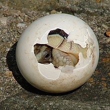
| A baby tortoise emerges from an amniotic egg | |
| Scientific classification | |
| Missing taxonomy template (fix): | Amniote |
| Clades | |
The amniotes are a group of tetrapods (four-limbed animals with backbones or spinal columns) that have a terrestrially adapted egg. They include synapsids (mammals along with their extinct kin) and sauropsids (reptiles and birds), as well as their fossil ancestors. Amniote embryos, whether laid as eggs or carried by the female, are protected and aided by several extensive membranes. In eutherian mammals (such as humans), these membranes include the amniotic sac that surrounds the fetus. These embryonic membranes, and the lack of a larval stage, distinguish amniotes from tetrapod amphibians.[1]
The first amniotes (referred to as "basal amniotes"), such as Casineria, resembled small lizards and had evolved from the amphibian reptiliomorphs about 340 million years ago, in the Carboniferous geologic period. Their eggs could survive out of the water, allowing amniotes to branch out into drier environments. The eggs could also "breathe" and cope with waste, allowing the eggs and the amniotes themselves to evolve into larger forms. The amniotes spread across the globe and became the dominant land vertebrates.
Very early in the evolutionary history of amniotes, basal amniotes evolved into two main lines of amniotes, the synapsids and the sauropsids, both of which persist into the modern era. The oldest known fossil synapsid is Protoclepsydrops from about 320 million years ago, while the oldest known sauropsid is probably Paleothyris, in the order Captorhinida, from the Middle Pennsylvanian epoch (ca. 306-312 million years ago).
Description
Amniotes can be characterized in part by embryonic development that includes the formation of several extensive membranes, the amnion, chorion, and allantois. Amniotes develop directly into a (typically) terrestrial form with limbs and a thick stratified epithelium, rather than first entering a feeding larval tadpole stage followed by metamorphosis as in amphibians. In amniotes the transition from a two-layered periderm to cornified epithelium is triggered by thyroid hormone during embryonic development, rather than metamorphosis.[2] The unique embryonic features of amniotes may reflect specializations of eggs to survive drier environments, or the massive size and yolk content of eggs evolved for direct development to a larger size.

1. Eggshell
2. Outer membrane
3. Inner membrane
4. Chalaza
5. Exterior albumen (outer thin albumen)
6. Middle albumen (inner thick albumen)
7. Vitelline membrane
8. Nucleus of Pander
9. Germinal disk (blastoderm)
10. Yellow yolk
11. White yolk
12. Internal albumen
13. Chalaza
14. Air cell
15. Cuticula
Adaptions for a terrestrial life
Features of amniotes evolved for survival on land include a sturdy but porous leathery or hard eggshell and an allantois evolved to facilitate respiration while providing a reservoir for disposal of wastes. Their kidneys and large intestines are also well-suited to water retention. Most mammals do not lay eggs, but corresponding structures may be found inside the placenta.
The first amniotes, such as Casineria kiddi, which lived about 340 million years ago, evolved from amphibian reptiliomorphs and resembled small lizards. Their eggs were small and covered with a leathery membrane, not a hard shell like those of birds or crocodiles. Although some modern amphibians lay eggs on land, with or without significant protection, they all lack advanced traits like an amnion. This kind of egg only became possible with internal fertilization. The outer membrane, a soft shell, evolved as a protection against the harsher environments on land, as species evolved to lay their eggs on land where they were safer than in the water. One can assume the ancestors of the amniotes laid their eggs in moist places, as such modest-sized animals would not have difficulty finding depressions under fallen logs or other suitable places in the ancient forests, and dry conditions were probably not the main reason why the soft shell emerged.[3] Indeed, many modern day amniotes are dependent on moisture to stop their eggs from desiccating.[4]
The egg membranes
In fish and amphibians there is only one inner membrane, also called an embryonic membrane. In amniotes the inner anatomy of the egg has evolved further and new structures have developed to take care of the gas exchanges between the embryo and the atmosphere, as well as dealing with the waste problems. To grow a thicker and tougher shell required new ways to supply the embryo with oxygen, as diffusion alone was not enough. After the egg developed these structures, further sophistication allowed amniotes to lay much bigger eggs in much drier habitats. Bigger eggs allowed for bigger offspring, and bigger adults could produce bigger eggs, so amniotes grew bigger than their ancestors. Real growth was not possible, however, until they stopped relying on small invertebrates as their main food source and started to eat plants or other vertebrates, or returned to the water.[dubious – discuss] New habits and heavier bodies meant further evolution for the amniotes, both in behavior and anatomy.
Amniote traits
While the early amniotes resembled their amphibian ancestors in many respects, a key difference was the lack of an otic notch at the back margin of the skull roof. In their ancestors, this notch held a spiracle, an unnecessary structure in an animal without an aquatic larval stage.[5] There are three main lines of amniotes, which may be distinguished by the structure of the skull and in particular the number of temporal fenestrae (openings) behind each eye. In anapsids (turtles) there are none, in synapsids (mammals and their extinct relatives) there is one, and in most diapsids (non-anapsid reptiles, including dinosaurs and birds) there are two.[6]
Post cranial remains of amniotes can be identified from their Labyrinthodont ancestors by their having at least two pairs of sacral ribs, a sternum in the pectoral girdle (some amniotes have lost it) and an astragalus bone in the ankle.[7]
Definition and classification
Amniota was first formally described by embryologist Ernst Haeckel in 1866 on the presence of the amnion, hence the name. A problem with this definition is that the trait (apomorphy) in question does not fossilize, and the status of fossil forms has to be inferred from other traits. Thus Jacques Gauthier and colleagues forwarded a definition of Amniota in 1988 as "the most recent common ancestor of extant mammals and reptiles, and all its descendants".[7] Gauthiers definition being a node-based crown group, his definition of the group has a slightly different content than the group defined as biological amniotes (apomorphy-based clade).[8]
Traditional classification
Classifications of the amniotes have traditionally recognised three classes based on major traits and physiology:[6][9][10][11]
- Class Reptilia (reptiles)
- Class Aves (birds)
- Subclass Neornithes (all modern birds, several extinct subclasses recognised)
- Class Mammalia (mammals)
- Subclass Monotremata (egg-laying mammals)
- Subclass Theria (marsupials and placental mammals)
This rather orderly scheme is the one most commonly found in popular and basic scientific works. It has come under critique from cladistics, as the class Reptilia is paraphyletic, that is, it has given rise to two other classes not included in Reptilia.
Phylogenetic classification
With the advent of cladistics, some researchers have attempted to establish new classes, based on phylogeny, but disregarding the physiological and anatomical unity of the groups. One such classification, by Michael Benton, is presented in simplified form below.[12]
- Clade Amniota ("reptiles")
- Class Synapsida - includes mammal-like reptiles
- Paraphyletic Order Pelycosauria †
- Order Therapsida
- Class Mammalia - Mammals
- Class Sauropsida
- Subclass Anapsida
- Order Testudines - Turtles
- Subclass Diapsida
- Order Araeoscelidia †
- Order Younginiformes †
- Infraclass Ichthyosauria †
- Infraclass Lepidosauromorpha
- Superorder Sauropterygia †
- Order Placodontia †
- Order Nothosauroidea †
- Order Plesiosauria †
- Superorder Lepidosauria
- Order Sphenodontida - Tuatara
- Order Squamata - Lizards & snakes
- Superorder Sauropterygia †
- Infraclass Archosauromorpha
- Order Prolacertiformes †
- Division Archosauria
- Subdivision Crurotarsi
- Order Crocodylia - Crocodilians
- Subdivision Avemetatarsalia
- Order Pterosauria †
- Superorder Dinosauria
- Order Ornithischia †
- Order Saurischia
- Class Aves - Birds
- Subdivision Crurotarsi
- Subclass Anapsida
- Class Synapsida - includes mammal-like reptiles
Cladogram
The cladogram presented here illustrates the phylogeny (family tree) of amniotes, and follows a simplified version of the relationships found by Laurin & Reisz (1995).[13] The cladogram covers the group as defined under Gauthier's definition.
| Amniota |
| ||||||||||||||||||||||||||||||
The inclusion of Testudines within Parareptilia is controversial. There is evidence from recent molecular studies that this group belongs in the Diapsida.
References
- ^ Benton, Michael J. (1997). Vertebrate Palaeontology. London: Chapman & Hall. pp. 105–109. ISBN 0-412-73810-4.
- ^ Alexander M. Schreiber and Donald D. Brown *. "Tadpole skin dies autonomously in response to thyroid hormone at metamorphosis". Pnas.org. Retrieved 2011-04-25.
- ^ Stewart J. R. (1997): Morphology and evolution of the egg of oviparous amniotes. In: S. Sumida and K. Martin (ed.) Amniote Origins-Completing the Transition to Land (1): 291-326. London: Academic Press.
- ^ Cunningham, B. (1938). "Further Studies on Water Absorption by Reptile Eggs". The American Naturalist. 72 (741): 380–385. doi:10.1086/280791. JSTOR 2457547.
{{cite journal}}: Unknown parameter|coauthors=ignored (|author=suggested) (help); Unknown parameter|month=ignored (help) - ^ Lombard, R. E. & Bolt, J. R. (1979): Evolution of the tetrapod ear: an analysis and reinterpretation. Biological Journal of the Linnean Society No 11: pp 19–76 Abstract
- ^ a b Romer, A.S. & Parsons, T.S. (1985): The Vertebrate Body. (6th ed.) Saunders, Philadelphia.
- ^ a b Gauthier, J., Kluge, A.G. and Rowe, T. (1988). "The early evolution of the Amniota." Pp. 103-155 in Benton, M.J. (ed.), The phylogeny and classification of the tetrapods, Volume 1: amphibians, reptiles, birds. Oxford: Clarendon Press.
- ^ Lee, M.S.Y. & Spencer, P.S. (1997): Crown clades, key characters and taxonomic stability: when is an amniote not an amniote? In: Sumida S.S. & Martin K.L.M. (eds.) Amniote Origins: completing the transition to land. Academic Press, pp 61-84. Google books
- ^ Carroll, R. L. (1988), Vertebrate Paleontology and Evolution, WH Freeman & Co.
- ^ Hildebrand, M. & G. E. Goslow, Jr. Principal ill. Viola Hildebrand. (2001). Analysis of vertebrate structure. New York: Wiley. p. 429. ISBN 0-471-29505-1.
{{cite book}}: CS1 maint: multiple names: authors list (link) - ^ Colbert, E.H. & Morales, M. (2001): Colbert's Evolution of the Vertebrates: A History of the Backboned Animals Through Time. 4th edition. John Wiley & Sons, Inc, New York — ISBN 978-0-471-38461-8.
- ^ Benton, M.J. (2004). Vertebrate Paleontology. Blackwell Publishers. xii-452. ISBN 0-632-05614-2.
{{cite book}}: Unknown parameter|nopp=ignored (|no-pp=suggested) (help) - ^ Laurin, M. and Reisz, R.R. (1995). "A reevaluation of early amniote phylogeny." Zoological Journal of the Linnean Society, 113: 165-223.
This article needs additional citations for verification. (January 2009) |
