
Taste bud: Difference between revisions
Rumilofaniel (talk | contribs) m Corrected minor syntax error resulting in non-displayable infobox. |
NEUROtiker (talk | contribs) m This may belong in tongue but certainly not at the beginning of this article |
||
| Line 13: | Line 13: | ||
DorlandsPre = c_03 | |
DorlandsPre = c_03 | |
||
DorlandsSuf = 12205927 }} |
DorlandsSuf = 12205927 }} |
||
The tongue is a movable muscle organ that lays on the floor of your mouth. The tongue helps you swallow, taste,and talk. |
|||
'''Taste buds''' are small structures on the upper surface of the [[tongue]], [[soft palate]], upper [[esophagus]] and [[epiglottis]] that provide information about the [[taste]] of food being eaten. These structures are involved in detecting the five elements of taste perception: salty, sour, bitter, sweet, and umami (or savory). Via small openings in the tongue epithelium, called taste pores, parts of the food dissolved in [[saliva]] come into contact with the [[taste receptor]]s. These are located on top of the [[taste receptor cell]]s that constitute the taste buds. The taste receptor cells send information detected by clusters of various receptors and ion channels to the gustatory areas of the brain via the seventh, ninth and tenth cranial nerves. |
'''Taste buds''' are small structures on the upper surface of the [[tongue]], [[soft palate]], upper [[esophagus]] and [[epiglottis]] that provide information about the [[taste]] of food being eaten. These structures are involved in detecting the five elements of taste perception: salty, sour, bitter, sweet, and umami (or savory). Via small openings in the tongue epithelium, called taste pores, parts of the food dissolved in [[saliva]] come into contact with the [[taste receptor]]s. These are located on top of the [[taste receptor cell]]s that constitute the taste buds. The taste receptor cells send information detected by clusters of various receptors and ion channels to the gustatory areas of the brain via the seventh, ninth and tenth cranial nerves. |
||
Revision as of 20:43, 12 September 2009
| Taste buds | |
|---|---|
 Semidiagrammatic view of a portion of the mucous membrane of the tongue. Two fungiform papillæ are shown. On some of the filiform papillæ the epithelial prolongations stand erect, in one they are spread out, and in three they are folded in. | |
 The mouth cavity. The cheeks have been slit transversely and the tongue pulled forward. | |
| Details | |
| Identifiers | |
| Latin | caliculus gustatorius |
| MeSH | D013650 |
| NeuroLex ID | birnlex_4101 |
| TA98 | A15.4.00.002 |
| TA2 | 7037 |
| TH | H3.04.01.0.02116, H3.04.01.0.03013 |
| FMA | 54825 |
| Anatomical terminology | |
Taste buds are small structures on the upper surface of the tongue, soft palate, upper esophagus and epiglottis that provide information about the taste of food being eaten. These structures are involved in detecting the five elements of taste perception: salty, sour, bitter, sweet, and umami (or savory). Via small openings in the tongue epithelium, called taste pores, parts of the food dissolved in saliva come into contact with the taste receptors. These are located on top of the taste receptor cells that constitute the taste buds. The taste receptor cells send information detected by clusters of various receptors and ion channels to the gustatory areas of the brain via the seventh, ninth and tenth cranial nerves.
The human tongue has about 10,000 taste buds.[citation needed]
Types of papillae
The majority of taste buds on the tongue sit on raised protrusions of the tongue surface called papillae. There are four types of papillae present in the human tongue:
- Fungiform papillae - as the name suggests, these are slightly mushroom-shaped if looked at in longitudinal section. These are present mostly at the apex (tip) of the tongue, as well as at the sides. Innervated by facial nerve.
- Filiform papillae - these are thin, long papillae "V"-shaped cones that don't contain taste buds but are the most numerous. These papillae are mechanical and not involved in gustation. Characterized increased keratinization.
- Foliate papillae - these are ridges and grooves towards the posterior part of the tongue found on lateral margins. Innervated by facial nerve (anterior papillae) and glossopharyngeal nerve (posterior papillae).
- Circumvallate papillae - there are only about 3-14 of these papillae on most people, and they are present at the back of the oral part of the tongue. They are arranged in a circular-shaped row just in front of the sulcus terminalis of the tongue. They are associated with ducts of Von Ebner's glands. Innervated by the glossopharyngeal nerve.
It is known that there are five taste sensations:
- Sweet, Bitter, and Umami (now sometimes called Savory), which work with a signal through a G protein-coupled receptor.
- Salty and Sour, which work with ion channels.
Localization of taste and the human "tongue map"
Contrary to popular understanding that different tastes map to different areas of the tongue, taste qualities are found in all areas of the tongue, although some regions are more sensitive than others.[1][2]
The original "tongue map" was based on a mistranslation by Harvard psychologist Edwin G. Boring of a German paper that was written in 1901.[3] Varying sensitivity to all tastes occurs across the whole tongue and indeed to other regions of the mouth where there are taste buds (epiglottis, soft palate).[4]
Structure of taste buds
Each taste bud is flask-like in shape, its broad base resting on the corium, and its neck opening, the gustatory pore, between the cells of the epithelium.
The bud is formed by two kinds of cells: supporting cells and gustatory cells.
- The supporting (sustentacular) cells are mostly arranged like the staves of a cask, and form an outer envelope for the bud. Some, however, are found in the interior of the bud between the gustatory cells.
- The gustatory (taste) cells, a chemoreceptor, occupy the central portion of the bud; they are spindle-shaped, and each possesses a large spherical nucleus near the middle of the cell.
The peripheral end of the cell terminates at the gustatory pore in a fine hair-like filament, the gustatory hair.
The central process passes toward the deep extremity of the bud, and there ends in single or bifurcated varicosities.
The nerve fibrils after losing their medullary sheaths enter the taste bud, and end in fine extremities between the gustatory cells; other nerve fibrils ramify between the supporting cells and terminate in fine extremities; these, however, are believed to be nerves of ordinary sensation and not gustatory.
See also
Additional images
-
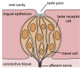 schematic drawing of a taste bud
 -
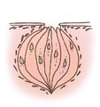 taste bud (nerve missing)


References
- ^ Huang A. L., et al. ""The cells and logic for mammalian sour taste detection""., Nature, 442. 934 - 938 (2006).
- ^ Scenta. "How sour taste buds grow". Retrieved August 28 2006.
{{cite web}}: Check date values in:|accessdate=(help); Unknown parameter|dateformat=ignored (help) - ^ Hänig, D.P., 1901. Zur Psychophysik des Geschmackssinnes. Philosophische Studien, 17: 576-623.
- ^ Collings, V.B., 1974. Human Taste Response as a Function of Locus of Stimulation on the Tongue and Soft Palate. Perception & Psychophysics, 16: 169-174.
External links
- Taste Perception: Cracking the Code
- Scientists Explore the Workings of Taste Buds from National Public Radio's Talk of the Nation, July 22, 2005