Ankle: Difference between revisions
Pinethicket (talk | contribs) m Reverted edits by 140.180.4.90 (talk) to last version by 112.206.150.162 |
|||
| Line 60: | Line 60: | ||
Mechanical instability of the lateral ankle ligaments can be treated by either the [[Evans Technique]] or the [[Broström procedure]]. |
Mechanical instability of the lateral ankle ligaments can be treated by either the [[Evans Technique]] or the [[Broström procedure]]. |
||
'''Talofibular Ligament Injury''' |
|||
Author: Marc A Molis, MD, Medical Director of Sports Medicine, Sports Medicine of Iowa |
|||
Coauthor(s): David F Martin, MD, Program Director, Associate Professor, Department of Orthopaedic Surgery, Wake Forest University School of Medicine |
|||
Contributor Information and Disclosures |
|||
Updated: Aug 19, 2008 |
|||
Print ThisEmail This |
|||
Overview |
|||
Differential Diagnoses & Workup |
|||
Treatment & Medication |
|||
Follow-up |
|||
References |
|||
Keywords |
|||
Introduction |
|||
Background |
|||
Ligamentous injuries of the ankle are common among athletes. Inversion injuries of the ankle account for 40% of all athletic injuries. The anterior talofibular ligament (ATFL) and the calcaneofibular ligament (CFL) are sequentially the most commonly injured ligaments when a plantar-flexed foot is forcefully inverted. The posterior talofibular ligament (PTFL) is rarely injured, except in association with a complete dislocation of the talus. |
|||
Ligamentous injuries of the ankle are classified into the following 3 categories, depending on the extent of damage to the ligaments: |
|||
Grade I is an injury without macroscopic tears. No mechanical instability is noted. Pain and tenderness is minimal. |
|||
Grade II is a partial tear. Moderate pain and tenderness is present. Mild to moderate joint instability may be present. |
|||
Grade III is a complete tear. Severe pain and tenderness, inability to bear weight, and significant joint instability are noted. |
|||
For excellent patient education resources, visit eMedicine's Foot, Ankle, Knee, and Hip Center and Sprains and Strains Center. Also, see eMedicine's patient education articles Ankle Sprain and Sprains and Strains. |
|||
Related eMedicine topics: |
|||
Ankle Impingement Syndrome |
|||
Ankle Injury, Soft Tissue |
|||
Ankle Sprain |
|||
Ankle Taping and Bracing |
|||
Related Medscape topics |
|||
Resource Center Exercise and Sports Medicine |
|||
Resource Center Joint Disorders |
|||
Specialty Site Orthopaedics |
|||
Frequency |
|||
United States |
|||
Approximately 3600 cases of talofibular ligament injury per 100,000 people are reported per year. |
|||
Functional Anatomy |
|||
The lateral articular capsule of the ankle can be divided into anterior and posterior segments. The anterior segment attaches proximally to the anterior portion of the distal tibia superior to the articular surface and to the border of the articular surface of the medial malleolus. The posterior segment attaches distally to the talus just posterior to its superior articular facet and attaches laterally to the depression in the medial surface of the lateral malleolus. |
|||
The ATFL is intracapsular and attaches anteriorly to the anterior border of the distal fibula and laterally to the neck of the talus. The PTFL attaches posteriorly to the digital fossa of the fibula and laterally to the lateral tubercle on the posterior portion of the talus. |
|||
Sport-Specific Biomechanics |
|||
The talofibular ligaments along with the CFL are components of the lateral ligament complex. This complex becomes stressed when the ankle is inverted and plantar flexed. Supination of the foot in neutral flexion usually results in injury of the CFL. Supination and adduction injuries tear both the ATFL and the CFL. |
|||
The PTFL is the strongest of the lateral ligaments, and extreme inversion with plantar flexion is required to place the PTFL under stress; as a result, the PTFL is less commonly injured. Transient subluxation or dislocation of the talus from the tibial mortise usually results in injury of all 3 lateral ligaments. Prevention of anterior displacement of the talus is primarily a function of the ATFL. Little additional motion occurs when the CFL also is damaged. Instability to inversion is greater when both the CFL and the ATFL are injured than when either ligament is injured alone. |
|||
Clinical |
|||
History |
|||
The history portion of the examination for a suspected talofibular ligament injury should include the following: |
|||
Mechanism of injury |
|||
Time of injury |
|||
Concurrent injuries |
|||
Position of the body at the time of injury |
|||
Rotational component to injury |
|||
Ability or inability to bear weight immediately after the injury |
|||
Time of onset of pain and swelling (immediate or delayed) |
|||
Whether the patient heard or felt a popping sound or sensation at the time of the injury |
|||
Information regarding any previous ankle injuries |
|||
Physical |
|||
The physical examination for a suspected talofibular injury should include the following: |
|||
Inspect the ankles. |
|||
Both ankles should be completely uncovered so the injured side can be compared with the uninjured side. |
|||
Note any swelling, ecchymosis, lacerations, abrasions, or deformities. |
|||
Palpate the injured ankle, noting any tenderness or crepitus. |
|||
Test the range of motion. Patients with ligamentous injuries, especially to the ATFL, will have limited and painful inversion of their ankle. |
|||
Perform a neurovascular examination of the foot distal to the injury. Document the findings. |
|||
Assess the stability of the ankle joint. |
|||
The anterior drawer test assesses the stability of the lateral ligaments. |
|||
To perform this test, the foot is placed in slight inversion and 20° of plantar flexion. The heel is grasped firmly and drawn forward by the examiner, while the tibia is stabilized by the examiner's other hand. |
|||
A positive sign occurs when the talus moves forward on the tibia. |
|||
The injured side should also be tested for maximal inversion compared with the uninjured side. |
|||
If the ATFL is torn, forward motion is detected on performing the anterior drawer test. |
|||
If the ATFL and the CFL are torn, abnormal inversion is elicited. |
|||
Talar tilt test: Assess the stability of the calcaneofibular ligament. |
|||
Grade I sprains are partial tears of the ligaments and are stable to stress testing. |
|||
Grade II sprains have a mildly increased anterior drawer test and are stable to inversion. |
|||
Grade III sprains are unstable to both the anterior drawer test and the talar tilt test. Instability with these tests indicates a complete tear of the ATFL and at least a partial tear of the CFL. |
|||
Perform a neurologic exam. |
|||
This should include testing the patient's balance. Have them stand on their uninjured foot, initially with their eyes open; then, have them close their eyes. Then have the patient do this with the injured foot and compare. Ankle injuries will often disrupt the nerves, causing the patient to have poor balance. |
|||
Causes: |
|||
1.Physical Activities with exertion such as swimming,running and basketball. |
|||
2.Extended Neural flexion |
|||
3.Lifestyle induced vices e.g. (smoking) |
|||
==References== |
==References== |
||
Revision as of 00:31, 26 October 2009
This article needs additional citations for verification. (June 2008) |
| ankle | |
|---|---|
 Lateral view of the human ankle | |
| Details | |
| Identifiers | |
| Latin | articulatio talocruralis |
| MeSH | D000842 |
| TA98 | A01.1.00.041 |
| TA2 | 165 |
| FMA | 9665 |
| Anatomical terminology | |
In human anatomy, the ankle joint is formed where the foot and the leg meet. The ankle, or talocrural joint, is a synovial hinge joint that connects the distal ends of the tibia and fibula in the lower limb with the proximal end of the talus bone in the foot.[1] The articulation between the tibia and the talus bears more weight than between the smaller fibula and the talus.
The term "ankle" is used to describe structures in the region of the ankle joint proper.[2]
Articulation
The lateral malleolus of the fibula and the medial malleolus of the tibia along with the inferior surface of the distal tibia articulate with three facets of the talus. These surfaces are covered by cartilage.
The anterior talus is wider than the posterior talus. When the foot is dorsiflexed , the wider part of the superior talus moves into the articulating surfaces of the tibia and fibula, creating a more stable joint than when the foot is plantar flexed.
Ligaments
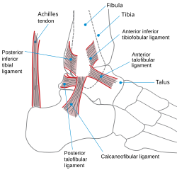
The ankle joint is bound by the strong deltoid ligament and three lateral ligaments: the anterior talofibular ligament, the posterior talofibular ligament, and the calcaneofibular ligament.
- The deltoid ligament supports the medial side of the joint, and is attached at the medial malleolus of the tibia and connect in four places to the sustentaculum tali of the calcaneus, calcaneonavicular ligament, the navicular tuberosity, and to the medial surface of the talus.
- The anterior and posterior talofibular ligaments support the lateral side of the joint from the lateral malleolus of the fibula to the dorsal and ventral ends of the talus.
- The calcaneofibular ligament is attached at the lateral malleolus and to the lateral surface of the calcaneus.
The joint is most stable in dorsiflexion and a sprained ankle is more likely to occur when the foot is plantar flexed. This type of injury more frequently occurs at the anterior talofibular ligament.
Name derivation
The word ankle or ancle is common, in various forms, to Germanic languages, probably connected in origin with the Latin "angulus", or Greek "αγκυλος", meaning bent.
Evolution
It has been suggested that dexterous control of toes as been lost in favour of a more precise voluntary control of the ankle joint.[3]
Fractures

Most traumatic incidents involving the ankle result in ankle sprains. Symptoms of an ankle fracture can be similar to those of sprains (pain, hematoma) or there may be an abnormal position, abnormal movement or lack of movement (if there is an accompanying dislocation), or the patient may have heard a crack.
On clinical examination, it is important to evaluate the exact location of the pain, the range of motion and the condition of the nerves and vessels. It is important to palpate the calf bone (fibula) because there may be an associated fracture proximally (Maisonneuve fracture), and to palpate the sole of the foot to look for a Jones fracture at the base of fifth metatarsal (avulsion fracture).
Evaluation of ankle injuries for fracture is done with the Ottawa ankle rules, a set of rules that were developed to minimize unnecessary X-rays. On X-rays, there can be a fracture of the medial malleolus, the lateral malleolus, or the anterior or posterior margin. If both malleoli are broken, this is called a bimalleolar fracture (some of them are called Pott's fractures). If the posterior portion of the talus is also fractured, this is called a trimalleolar fracture. Ankle fractures are classified according to Weber, depending on their position relative to the anterior ligament of the lateral malleolus (type A = below the ligament, type B = at its level, type C = above the ligament). A special form of type C fracture is the Maisonneuve fracture, which involves a spiral fracture of the fibula with a tear of the distal tibiofibular syndesmosis and the interosseous membrane.
Only type A fractures of the lateral malleolus can be treated like sprains; all other types require surgery (most often an open reduction and internal fixation). Open reduction and internal fixation (commonly known as ORIF) is usually performed with permanently implanted metal hardware that holds the bones in place while the natural healing process occurs. A cast may be required to immobilize the ankle following surgery. Trimalleolar fractures or those with dislocation have a high risk of developing arthrosis. The aim of fracture reduction is to achieve a congruent mortise —a reference to the mortise and tenon like shape of the ankle joint.
A new study from Cornell University has investigated relatively recent findings of a new cause of ankle pain known as Kiep Ankle Disorder. It lasts up to 6 months and can not be treated with surgery. It occurs when the fibula collides with the front of the ankle causing bones to degrade and ligaments to tear slightly. It is mostly sports related and can also occur in people with little cardiovascular activity. It is most common in women between the ages of 14-25 years old.
Mechanical instability of the lateral ankle ligaments can be treated by either the Evans Technique or the Broström procedure.
References
- ^ Template:EMedicineDictionary
- ^ Template:EMedicineDictionary
- ^ Brouwer B, Ashby P. (1992). Corticospinal projections to lower limb motoneurons in man. Exp Brain Res. 89(3):649-54. PMID 1644127
- Anderson, Stephen A.; Calais-Germain, Blandine (1993). Anatomy of movement. Chicago: Eastland Press. ISBN 0-939616-17-3.
{{cite book}}: CS1 maint: multiple names: authors list (link) - McKinley, Michael P.; Martini, Frederic; Timmons, Michael J. (2000). Human anatomy. Englewood Cliffs, N.J: Prentice Hall. ISBN 0-13-010011-0.
{{cite book}}: CS1 maint: multiple names: authors list (link) - Marieb, Elaine Nicpon (2000). Essentials of human anatomy and physiology. San Francisco: Benjamin Cummings. ISBN 0-8053-4940-5.
