5S ribosomal RNA: Difference between revisions
Appearance
Content deleted Content added
m remove dupe link |
Converting from rfambox to infobox rfam template |
||
| Line 1: | Line 1: | ||
{{Infobox rfam |
|||
{{Rfam_box|acc=RF00001| description=5S ribosomal RNA |abbreviation=5S_rRNA |avg_length=114.50 |avg_identity=59.00 |type=Gene; rRNA; |se=Szymanski et al, 5S ribosomal database, {{PMID|11752286}} |ss=Published; {{PMID|11283358}} |release=10.0}} |
|||
| Name = 5S ribosomal RNA |
|||
| image = RF00001.jpg |
|||
| width = |
|||
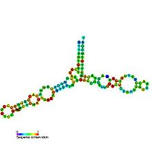
| caption = Predicted [[secondary structure]] and [[sequence conservation]] of 5S ribosomal RNA |
|||
| Symbol = 5S_rRNA |
|||
| AltSymbols = |
|||
| Rfam = RF00001 |
|||
| miRBase = |
|||
| miRBase_family = |
|||
| RNA_type = [[Gene]]; [[Ribosomal RNA | rRNA]]; |
|||
| Tax_domain = [[Eukaryota]];[[ Bacteria]];[[ Viruses]];[[ Archaea]]; |
|||
| GO = {{GO|0005840}} {{GO|0003735}} |
|||
| SO = {{SO|0000652}} |
|||
| CAS_number = |
|||
| EntrezGene = |
|||
| HGNCid = |
|||
| OMIM = |
|||
| PDB = |
|||
| RefSeq = |
|||
| Chromosome = |
|||
| Arm = |
|||
| Band = |
|||
| LocusSupplementaryData = |
|||
}} |
|||
[[Image:PDB 1c2x EBI.png|thumb|200px|left|A 3D representation of a 5S rRNA molecule. This structure is of the 5S rRNA from the Escherichia coli 50S ribosomal subunit and is based on a [[Transmission electron microscopy|cryo-electron microscopic reconstruction]].<ref name="pmid10756104">{{cite journal |author=Mueller F, Sommer I, Baranov P, Matadeen R, Stoldt M, Wöhnert J, Görlach M, van Heel M, Brimacombe R |title=The 3D arrangement of the 23 S and 5 S rRNA in the Escherichia coli 50 S ribosomal subunit based on a cryo-electron microscopic reconstruction at 7.5 A resolution. |journal=J Mol Biol |volume=298 |issue=1 |pages=35–59 |year=2000 |pmid=10756104 |doi=10.1006/jmbi.2000.3635}}</ref>]] |
[[Image:PDB 1c2x EBI.png|thumb|200px|left|A 3D representation of a 5S rRNA molecule. This structure is of the 5S rRNA from the Escherichia coli 50S ribosomal subunit and is based on a [[Transmission electron microscopy|cryo-electron microscopic reconstruction]].<ref name="pmid10756104">{{cite journal |author=Mueller F, Sommer I, Baranov P, Matadeen R, Stoldt M, Wöhnert J, Görlach M, van Heel M, Brimacombe R |title=The 3D arrangement of the 23 S and 5 S rRNA in the Escherichia coli 50 S ribosomal subunit based on a cryo-electron microscopic reconstruction at 7.5 A resolution. |journal=J Mol Biol |volume=298 |issue=1 |pages=35–59 |year=2000 |pmid=10756104 |doi=10.1006/jmbi.2000.3635}}</ref>]] |
||
Revision as of 15:16, 11 October 2011
| 5S ribosomal RNA | |
|---|---|
 Predicted secondary structure and sequence conservation of 5S ribosomal RNA | |
| Identifiers | |
| Symbol | 5S_rRNA |
| Rfam | RF00001 |
| Other data | |
| RNA type | Gene; rRNA; |
| Domain(s) | Eukaryota;Bacteria;Viruses;Archaea; |
| GO | GO:0005840 GO:0003735 |
| SO | SO:0000652 |
| PDB structures | PDBe |


5S ribosomal RNA (5S rRNA) is a component of the large ribosomal subunit in both prokaryotes (50S) and eukaryotes (60S).
Eukaryotic 5S rRNA is synthesised by RNA polymerase III, whereas most other eukaroytic rRNAs are cleaved from a 45S precursor transcribed by RNA polymerase I. In Xenopus oocytes, it has been shown that fingers 4-7 of the nine-zinc finger transcription factor TFIIIA can bind to the central region of 5S RNA.[3] Binding between 5S rRNA and TFIIIA serves to both repress further transcription of the 5S RNA gene and stabilize the 5S RNA transcript until it is required for ribosome assembly.[4]
References
- ^ Mueller F, Sommer I, Baranov P, Matadeen R, Stoldt M, Wöhnert J, Görlach M, van Heel M, Brimacombe R (2000). "The 3D arrangement of the 23 S and 5 S rRNA in the Escherichia coli 50 S ribosomal subunit based on a cryo-electron microscopic reconstruction at 7.5 A resolution". J Mol Biol. 298 (1): 35–59. doi:10.1006/jmbi.2000.3635. PMID 10756104.
{{cite journal}}: CS1 maint: multiple names: authors list (link) - ^ Mitra K, Schaffitzel C, Fabiola F, Chapman MS, Ban N, Frank J (2006). "Elongation arrest by SecM via a cascade of ribosomal RNA rearrangements". Mol Cell. 22 (4): 533–43. doi:10.1016/j.molcel.2006.05.003. PMID 16713583.
{{cite journal}}: CS1 maint: multiple names: authors list (link) - ^ Searles, MA (2000). "The role of the central zinc fingers of transcription factor IIIA in binding to 5 S RNA". J Mol Biol. 301 (1): 47–60. doi:10.1006/jmbi.2000.3946. PMID 10926492.
{{cite journal}}: Unknown parameter|coauthors=ignored (|author=suggested) (help) - ^ Pelham, HRB (1980). "A specific transcription factor that can bind either the 5S RNA gene or 5S RNA". Proc Natl Acad Sci. 77 (7): 4170–4174. doi:10.1073/pnas.77.7.4170. PMC 349792. PMID 7001457.
{{cite journal}}: Unknown parameter|coauthors=ignored (|author=suggested) (help)
External links
- Page for 5S ribosomal RNA at Rfam
- 5SData
- 5S+Ribosomal+RNA at the U.S. National Library of Medicine Medical Subject Headings (MeSH)
