
Aortic arch
| Aortic arch | |
|---|---|
 Plan of the branches. | |
 The arch of the aorta, and its branches. | |
| Details | |
| Source | ascending aorta |
| Branches | brachiocephalic artery, left common carotid artery, left subclavian artery |
| Identifiers | |
| Latin | arcus aortae |
| TA98 | A12.2.04.001 |
| TA2 | 4177 |
| FMA | 3768 |
| Anatomical terminology | |
- For the embryological structure, see Aortic arches.
The arch of the aorta (Transverse Aorta) begins at the level of the upper border of the second sternocostal articulation of the right side, and runs at first upward, backward, and to the left in front of the trachea; it is then directed backward on the left side of the trachea and finally passes downward on the left side of the body of the fourth thoracic vertebra, at the lower border of which it becomes continuous with the descending aorta.
It thus forms two curvatures: one with its convexity upward, the other with its convexity forward and to the left. Its upper border is usually about 2.5 cm. below the superior border to the manubrium sterni.
Relations
The arch of the aorta is covered anteriorly by the pleura and anterior margins of the lungs, and by the remains of the thymus.
As the vessel runs backward its left side is in contact with the left lung and pleura.
Passing downward on the left side of this part of the arch are four nerves; in order from before backward these are, the left phrenic, the lower of the superior cardiac branches of the left vagus, the superior cardiac branch of the left sympathetic, and the trunk of the left vagus.
As the left vagus nerve crosses the arch, it gives off its recurrent branch, which hooks around below the vessel and then passes upward on its right side.
The highest left intercostal vein runs obliquely upward and forward on the left side of the arch, between the phrenic and vagus nerves.
On the right are the deep part of the cardiac plexus, the left recurrent nerve, the esophagus, and the thoracic duct; the trachea lies behind and to the right of the vessel.
Above are the innominate, left common carotid, and left subclavian arteries, which arise from the convexity of the arch and are crossed close to their origins by the left innominate vein.
Below are the bifurcation of the pulmonary artery, the left bronchus, the ligamentum arteriosum, the superficial part of the cardiac plexus, and the left recurrent nerve.
As already stated, the ligamentum arteriosum connects the commencement of the left pulmonary artery to the aortic arch.
Additional images
-
 Diagram showing the origins of the main branches of the carotid arteries.
 -
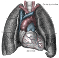 Front view of heart and lungs.
 -
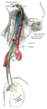 Course and distribution of the glossopharyngeal, vagus, and accessory nerves.


![]() This article incorporates text in the public domain from page 547 of the 20th edition of Gray's Anatomy (1918)
This article incorporates text in the public domain from page 547 of the 20th edition of Gray's Anatomy (1918)
{