
Esophagus
This article needs additional citations for verification. (December 2007) |
| Esophagus | |
|---|---|
 Head and neck. | |
 Digestive organs. (oEsophagus is #1) | |
| Details | |
| Precursor | Foregut |
| Artery | oesophageal arteries |
| Vein | oesophageal veins |
| Nerve | celiac ganglia, vagus[1] |
| Identifiers | |
| Latin | Oesophagus(NECK) |
| MeSH | D004947 |
| TA98 | A05.4.01.001 |
| TA2 | 2887 |
| FMA | 7131 |
| Anatomical terminology | |
The esophagus or oesophagus (see American and British English spelling differences), sometimes known as the gullet, is an organ in vertebrates which consists of a muscular tube through which food passes from the pharynx to the stomach. In humans the oesophagus is continuous with the laryngeal part of the pharynx at the level of the C6 vertebra. It is usually 25-30 cm long which connects the mouth to the stomach. It is divided into cervical, thoracic, and abdominal parts. The trachea branches off the esophagus to the lungs.[2]
Functions of the esophagus
Food is passed through the oesophagus by using the process of peristalsis. Specifically, it connects the pharynx, which is the body cavity that is common to the digestive factory and respiratory system with the stomach, where the second stage of digestion is initiated.
The oesophagus is lined with mucous membrane, and is more deeply lined with muscle that acts with peristaltic action to move swallowed food down to the stomach.
The swallowing sound that we hear is the esophagus functioning.
Histology
The layers of the esophagus are as follows:[3]
- mucosa
- nonkeratinized stratified squamous epithelium: is rapidly turned over, and serves a protective effect due to the high volume transit of food, saliva and mucous.
- lamina propria: sparse.
- muscularis mucosae: smooth muscle
- submucosa: Contains the mucous secreting glands (esophageal glands), and connective structures termed papillae.
- muscularis externa (or "muscularis propria"): composition varies in different parts of the oesophagus, to correspond with the conscious control over swallowing in the upper portions and the autonomic control in the lower portions:
- upper third, or superior part: striated muscle
- middle third, smooth muscle and striated muscle,
- inferior third: predominantly smooth muscle.
- adventitia
Gastroesophageal junction
The junction between the esophagus and the stomach (the gastroesophageal junction or GE junction) is not actually considered a valve, although it is sometimes called the cardiac sphincter, cardia or cardias, but is actually more of a stricture.
Esophageal diseases and conditions
Many people experience a burning sensation in their chest occasionally, caused by stomach acids refluxing into the oesophagus, normally called heartburn. Extended exposure to heartburn may erode the lining of the oesophagus, leading potentially to Barrett's oesophagus which is associated an increased risk of adenocarcinoma most commonly found in the distal one-third of the oesophagus.
Some people also experience a sensation known as globus oesophagus, where it feels as if a ball is lodged in the lower part of the oesophagus.
The following are additional diseases and conditions that affect the oesophagus:
- Achalasia
- Chagas disease
- Caustic injury to the oesophagus
- Esophageal atresia and Tracheoesophageal fistula
- Esophageal cancer
- Esophageal web
- Esophagitis
- GERD
- Hiatus hernia
- Mallory-Weiss syndrome
- Neurogenic dysphagia
- Schatzki's ring
- Zenker's Diverticulum
- Boerhaave syndrome
Additional images
-
 Layers of the esophagus.
 -
 Mid-esophageal mass
 -
 Stomach
 -
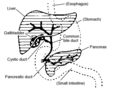 Accessory digestive system.
 -
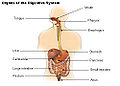 Organs of the digestive tract.
 -
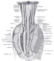
 Section of the neck at about the level of the sixth cervical vertebra.
 -
 Transverse section of thorax, showing relations of pulmonary artery.
 -
 Sagittal section of nose mouth, pharynx, and larynx.
 -
-
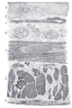 Section of the human oesophagus. Moderately magnified.
 -
 Microscopic shot of a cross section of human gastro-esophageal junction wall.


References
- ^ Template:GeorgiaPhysiology
- ^ Maton, Anthea (1993). Human Biology and Health. Englewood Cliffs, New Jersey, USA: Prentice Hall. ISBN 0-13-981176-1.
{{cite book}}: Unknown parameter|coauthors=ignored (|author=suggested) (help) - ^ Histology image: 10801loa – Histology Learning System at Boston University