
Thoracic diaphragm
| diaphragm | |
|---|---|
 Respiratory system | |
| Details | |
| Precursor | septum transversum, pleuroperitoneal folds, body wall [1] |
| Artery | Pericardiacophrenic artery, Musculophrenic artery, Inferior phrenic arteries |
| Vein | Superior phrenic vein, Inferior phrenic vein |
| Nerve | phrenic and lower intercostal nerves |
| Actions | respiration |
| Identifiers | |
| Latin | diaphragma |
| MeSH | D003964 |
| TA98 | A04.4.02.001 |
| TA2 | 2327 |
| FMA | 13295 |
| Anatomical terminology | |
- For other types of diaphragm, see Diaphragm.
In the anatomy of mammals, the thoracic diaphragm is a sheet of muscle extending across the bottom of the ribcage. The diaphragm separates the thoracic cavity from the abdominal cavity and performs an important function in respiration. A diaphragm in anatomy can refer to other flat structures such as the urogenital diaphragm or pelvic diaphragm, but "the diaphragm" generally refers to the thoracic diaphragm. Other vertebrates such as amphibians and reptiles have diaphragms or diaphragm-like structures, but important details of the anatomy vary, such as the position of lungs in the abdominal cavity.
Function
The diaphragm is crucial for breathing and respiration. During inhalation, the diaphragm contracts, thus enlarging the thoracic cavity (the external intercostal muscles also participate in this enlargement). This reduces intra-thoracic pressure: In other words, enlarging the cavity creates suction that draws air into the lungs. When the diaphragm relaxes, air is exhaled by elastic recoil of the lung and the tissues lining the thoracic cavity in conjunction with the abdominal muscles, which act as an antagonist paired with the diaphragm's contraction.
It is not responsible for all the breathing related to voice, a common misconception espoused by many teachers but few great singers. One has more control over the abdominals and intercostals than the actual diaphragm, which has relatively few proprioceptive nerve endings. By training proper posture and balance in the rest of the body, the diaphragm naturally strengthens and works in concert with surrounding structures rather than in isolation.
The diaphragm is also involved in non-respiratory functions, helping to expel vomit, feces, and urine from the body by increasing intra-abdominal pressure, and preventing acid reflux by exerting pressure on the esophagus as it passes through the esophageal hiatus.
In veterinary anatomy, the diaphragm is not necessarily crucial; a cow, for instance, can survive fairly asymptomatically with diaphragmatic paralysis as long as no massive aerobic metabolic demands are made of her.
Anatomy
The Diaphragm is a dome-shaped musculofibrous septum that separates the thoracic from the abdominal cavity, its convex upper surface forming the floor of the former, and its concave under surface the roof of the latter. Its peripheral part consists of muscular fibers that take origin from the circumference of the thoracic outlet and converge to be inserted into a central tendon.
The muscular fibers may be grouped according to their origins into three parts:
| Part | Origin |
| sternal | two fleshy slips from the back of the xiphoid process. |
| costal | the inner surfaces of the cartilages and adjacent portions of the lower six ribs on either side, interdigitating with the Transversus abdominis. |
| lumbar | aponeurotic arches, named the lumbocostal arches, and from the lumbar vertebrae by two pillars or crura. |
There are two lumbocostal arches, a medial and a lateral, on either side.
Innervation
The diaphragm is innervated by the phrenic nerve. It's a branch of C3,C4,and C5.
- you can remember that by "3, 4, 5 keeps the diaphragm alive"
Crura and central tendon
At their origins the crura are tendinous in structure, and blend with the anterior longitudinal ligament of the vertebral column.
The central tendon of the diaphragm is a thin but strong aponeurosis situated near the center of the vault formed by the muscle, but somewhat closer to the front than to the back of the thorax, so that the posterior muscular fibers are the longer.
Openings in the Diaphragm

The diaphragm is pierced by a series of apertures to permit of the passage of structures between the thorax and abdomen. Three large openings — the aortic, the esophageal, and the vena cava — and a series of smaller ones are described.
| opening | level | structures |
| caval opening | T8 | inferior vena cava, and some branches of the right phrenic nerve |
| esophageal hiatus | T10 | esophagus, the anterior and posterior vagal trunks, and some small esophageal arteries |
| aortic hiatus | T12 | the aorta, the azygos vein, and the thoracic duct |
| two lesser aperture of right crus | greater and lesser right splanchnic nerves | |
| three lesser aperture of left crus | greater and lesser left splanchnic nerves and the hemiazygos vein | |
| behind the diaphragm, under the medial lumbocostal arches | sympathetic trunk | |
| areolar tissue between the sternal and costal parts (see also foramina of Morgagni) | the superior epigastric branch of the internal mammary artery and some lymphatics from the abdominal wall and convex surface of the liver | |
| areolar tissue between the fibers springing from the medial and lateral lumbocostal arches | This interval is less constant; when this interval exists, the upper and back part of the kidney is separated from the pleura by areolar tissue only. |

You can remember some of them by this Mnemonic
- Aortic hiatus = 12 letters = T12
- Oesophagus = 10 letters = T10
- Vena cava = 8 letters = T8
Comparative anatomy and evolution
The existence of some membrane separating the pharynx from the stomach can be traced widely among the chordates. Thus amphioxus possesses an atrium by which water exits the pharynx, which has been argued (and disputed) to be homologous to structures in ascidians and hagfishes.[3] The urochordate epicardium separates digestive organs from the pharynx and heart, but the anus returns to the upper compartment to discharge wastes through an outgoing siphon.
Thus the diaphragm emerges in the context of a body plan that separated an upper feeding compartment from a lower digestive tract, but the point at which it originates is a matter of definition. Structures in fish, amphibians, reptiles, and birds have been called diaphragms, but it has been argued that these structures are not homologous. For instance, the alligator diaphragmaticus muscle does not insert on the esophagus and does not affect pressure of the lower esophageal sphincter.[4] The lungs are located in the abdominal compartment of amphibians and reptiles, so that contraction of the diaphragm expels air from the lungs rather than drawing it into them. In birds and mammals, lungs are located above the diaphragm. The presence of an exceptionally well-preserved fossil of Sinosauropteryx, with lungs located beneath the diaphragm as in crocodiles, has been used to argue that dinosaurs could not have sustained an active warm-blooded physiology, or that birds could not have evolved from dinosaurs.[5] An explanation for this state of affairs is that, when lungs originated beneath the diaphragm, but as the demands for respiration increased in warm-blooded birds and mammals, natural selection came to favor the parallel evolution of the herniation of the lungs from the abdominal cavity in both lineages.[2]. However, birds do not have diaphragms. They do not breathe in the same way as mammals, and do not rely on creating a negative pressure in the thoracic cavity, at least not to the same extent. They rely on a rocking motion of the keel of the sternum to create local areas of reduced pressure to supply thin, membranous airsacs cranially and caudally to the fixed-volume, non-expansive lungs. A complicated system of valves and air sacs cycles air constantly over the absorption surfaces of the lungs so allowing maximal efficiency of gaseous exchange. Thus, birds do not have the reciprocal tidal breathing flow of mammals. On careful dissection, around eight air sacs can be clearly seen. They extend quite far caudally into the abdomen. [6]
Variations
The sternal portion of the muscle is sometimes wanting and more rarely defects occur in the lateral part of the central tendon or adjoining muscle fibers.

CONTENTS · INDEX · ILLUSTRATIONS · BIBLIOGRAPHIC RECORD
  The American Heritage® Dictionary of the English Language: Fourth Edition.  2000.
 rayah
SYLLABICATION: ra·yah PRONUNCIATION: räy, r VARIANT FORMS: also ra·ya NOUN: A Christian subject under an Ottoman ruler. ETYMOLOGY: Turkish râya, from Arabic ra‘yah, subject, flock, from ra‘, to pasture, feed. See rcy in Appendix II.
The American Heritage® Dictionary of the English Language, Fourth Edition. Copyright © 2000 by Houghton Mifflin Company. Published by the Houghton Mifflin Company. All rights reserved.
Ray, Satyajit Rayburn, Samuel Taliaferro
Pathology
The right crus of the diaphragm is part of the lower esophageal sphincter (LES), which separates the thoracic and abdominal parts of the esophagus. A hiatal hernia, in which the abdominal esophagus or even the fundus of the stomach rises up through the esophageal hiatus into the thoracic cavity, can result from a tear or weakness in the diaphragm. Both general weakness of the LES and hiatal hernia can cause "acid reflex," also known as gastroesophageal reflex disease (GERD).
If the diaphragm is struck, or otherwise spasms, breathing will become difficult. This is called "having the wind knocked out of you."
A hiccup occurs when the diaphragm contracts periodically without voluntary control.
Diaphragmatic injuries result from either blunt or penetrating trauma.
Gas under the diaphragm may be pneumoperitoneum. CONTENTS · INDEX · ILLUSTRATIONS · BIBLIOGRAPHIC RECORD
The American Heritage® Dictionary of the English Language: Fourth Edition. 2000. rayah
SYLLABICATION: ra·yah PRONUNCIATION: räy, r VARIANT FORMS: also ra·ya NOUN: A Christian subject under an Ottoman ruler. ETYMOLOGY: Turkish râya, from Arabic ra‘yah, subject, flock, from ra‘, to pasture, feed. See rcy in Appendix II.
The American Heritage® Dictionary of the English Language, Fourth Edition. Copyright © 2000 by Houghton Mifflin Company. Published by the Houghton Mifflin Company. All rights reserved.
CONTENTS · INDEX · ILLUSTRATIONS · BIBLIOGRAPHIC RECORD
Additional images
-
 The phrenic nerve and its relations with the vagus nerve.
 -
 Plan of right sympathetic cord and splanchnic nerves.
 -
 Thoracic portion of the sympathetic trunk.
 -
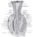 The position and relation of the esophagus in the cervical region and in the posterior mediastinum. Seen from behind.
 -
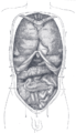 Front view of the thoracic and abdominal viscera.
 -
 The duodenum and pancreas.
 -
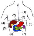
 Topography of thoracic and abdominal viscera.
 -
 Organs of abdomen
 -
 Peritoneopericardial diaphragmatic hernia in a dog.
 -

-
 Probably the first dictionary definition of "diaphragm"


CONTENTS · INDEX · ILLUSTRATIONS · BIBLIOGRAPHIC RECORD
rayah
SYLLABICATION: ra·yah PRONUNCIATION: räy, r VARIANT FORMS: also ra·ya NOUN: A Christian subject under an Ottoman ruler. ETYMOLOGY: Turkish râya, from Arabic ra‘yah, subject, flock, from ra‘, to pasture, feed. See rcy in Appendix II.
The American Heritage® Dictionary of the English Language, Fourth Edition. Copyright © 2000 by Houghton Mifflin Company. Published by the Houghton Mifflin Company. All rights reserved.
CONTENTS · INDEX · ILLUSTRATIONS · BIBLIOGRAPHIC RECORD
See also
References
- ^ mslimb-012—Embryo Images at University of North Carolina
- ^ a b Arthur Keith, M.D. (1905). "The nature of the mammalian diaphragm and pleural cavities". Journal of Anatomy and Physiology.
- ^ Zbynek Kozmik; et al. (1999). "Characterization of an amphioxus paired box gene, AmphiPax2/5/8" (PDF). Development. pp. 1295–1304.
{{cite web}}: Explicit use of et al. in:|author=(help) - ^ T. J. Uriona; et al. (2005). "Structure and function of the esophagus of the American alligator (Alligator mississippiensis)". Journal of Experimental Biology. pp. 3047–3053.
{{cite web}}: Explicit use of et al. in:|author=(help) - ^ "Lung fossils suggest that dinos breathed in cold blood".
- ^ Dyce, Sack and Wensing in Textbook of Veterinary Anatomy; 2002 (3rd Edn); Saunders, Philiadelphia
![]() This article incorporates text from a publication now in the public domain: Chambers, Ephraim, ed. (1728). Cyclopædia, or an Universal Dictionary of Arts and Sciences (1st ed.). James and John Knapton, et al.
This article incorporates text from a publication now in the public domain: Chambers, Ephraim, ed. (1728). Cyclopædia, or an Universal Dictionary of Arts and Sciences (1st ed.). James and John Knapton, et al. {{cite encyclopedia}}: Missing or empty |title= (help)
External links
- . GPnotebook https://www.gpnotebook.co.uk/simplepage.cfm?ID=483393594.
{{cite web}}: Missing or empty|title=(help) - Template:MuscleLoyola
![]() This article incorporates text in the public domain from page 404 of the 20th edition of Gray's Anatomy (1918)
This article incorporates text in the public domain from page 404 of the 20th edition of Gray's Anatomy (1918)