
Lateral rotator group
| Lateral rotator group | |
|---|---|
 The lateral rotator group and the gluteus minimus muscle | |
 Structures surrounding right hip joint | |
| Details | |
| Origin | At or below the acetabulum of the ilium |
| Insertion | On or near the greater trochanter of the femur |
| Artery | Inferior gluteal artery, lateral sacral artery, superior gluteal artery |
| Nerve | Obturator nerve, nerve to the Piriformis, nerve to quadratus femoris |
| Actions | Lateral rotation of hip |
| Antagonist | Gluteus minimus muscle, gluteus medius muscle |
| Anatomical terms of muscle | |
The lateral rotator group is a group of six small muscles of the hip which all externally (laterally) rotate the femur in the hip joint. It consists of the following muscles: Piriformis, gemellus superior, obturator internus, gemellus inferior, quadratus femoris and the obturator externus.[1]
All muscles in the lateral rotator group originate from the hip bone and insert on to the upper extremity of the femur. The muscles are innervated by the sacral plexus (L4-S2), except the obturator externus, which is innervated by the lumbar plexus.[2]
Individual muscles
| Muscle | origin | insertion | innervation[2] |
|---|---|---|---|
| Piriformis muscle | Anterior surface of sacrum between and laterally to the anterior sacral foramina | Superior boundary of greater trochanter | Nerve to the piriformis (S1-S2) |
| Gemellus superior muscle | Ischial spine | Upper edge of Obturator internus muscle tendon (indirectly greater trochanter) | Nerve to obturator internus (L5-S2) |
| Obturator internus muscle | Medial surface of obturator membrane and the surrounding bone | Medial surface of greater trochanter | Nerve to obturator internus (L5-S2) |
| Gemellus inferior muscle | Just above the tuberosity of the ischium | Lower edge of Obturator internus muscle tendon (indirectly greater trochanter) | Nerve to quadratus femoris (L4-S1) |
| Quadratus femoris muscle | Lateral edge of the tuberosity of the ischium | Intertrochanteric crest | Nerve to quadratus femoris (L4-S1) |
| Obturator externus muscle | Lateral surface of obturator membrane and the ischiopubic ramus | Trochanteric fossa | Posterior branch of obturator nerve (L3-L4) |
Other lateral rotators
This group does not include all muscles which aid in lateral rotation of the hip joint: rather it is a collection of ones which are known for primarily performing this action. Other muscles that contribute to lateral rotation of the hip include:
- Gluteus maximus muscle (lower fibres)
- Gluteus medius muscle and gluteus minimus muscle when the hip is extended (become medial rotators when hip is flexed)
- Psoas major muscle
- Psoas minor muscle
- Sartorius muscle
Additional images
-
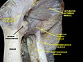
 Dissection of lateral rotator group (obturator externus hidden under quadratus femoris muscle) seen from the back
 -
 Muscles of thigh as seen from the front
 -
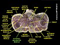 Cross section of pelvic region


See also
References
- ^ MedicalMnemonics.com: 833 3471 657
- ^ a b Bojsen-Møller, Finn; Simonsen, Erik B.; Tranum-Jensen, Jørgen (2001). Bevægeapparatets anatomi (in Danish) (12th ed.). p. 365. ISBN 978-87-628-0307-7.
{{cite book}}: Unknown parameter|trans_title=ignored (|trans-title=suggested) (help)
External links
- Glutealregion at The Anatomy Lesson by Wesley Norman (Georgetown University)