OSER1
| OSER1 | |||||||||||||||||||||||||||||||||||||||||||||||||||
|---|---|---|---|---|---|---|---|---|---|---|---|---|---|---|---|---|---|---|---|---|---|---|---|---|---|---|---|---|---|---|---|---|---|---|---|---|---|---|---|---|---|---|---|---|---|---|---|---|---|---|---|
| Identifiers | |||||||||||||||||||||||||||||||||||||||||||||||||||
| Aliases | OSER1, C20orf111, HSPC207, Osr1, Perit1, dJ1183I21.1, oxidative stress responsive serine rich 1 | ||||||||||||||||||||||||||||||||||||||||||||||||||
| External IDs | MGI: 1913930; HomoloGene: 9521; GeneCards: OSER1; OMA:OSER1 - orthologs | ||||||||||||||||||||||||||||||||||||||||||||||||||
| |||||||||||||||||||||||||||||||||||||||||||||||||||
| |||||||||||||||||||||||||||||||||||||||||||||||||||
| |||||||||||||||||||||||||||||||||||||||||||||||||||
| |||||||||||||||||||||||||||||||||||||||||||||||||||
| Wikidata | |||||||||||||||||||||||||||||||||||||||||||||||||||
| |||||||||||||||||||||||||||||||||||||||||||||||||||
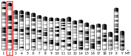
OSER1 (Oxidative Stress Responsive Serine Rich 1), or Chromosome 20 open reading frame 111, C20orf111, is the hypothetical protein that in humans is encoded by the OSER1 gene.[5] OSER1/C20orf111 is also known as Perit1 (peroxide inducible transcript 1), HSPC207, and dJ1183I21.1.[6] It was originally located using genomic sequencing of chromosome 20.[7] The National Center for Biotechnology Information, or NCBI,[5] shows that it is located at q13.11 on chromosome 20, however the genome browser at the University of California-Santa Cruz (UCSC) website[8] shows that it is at location q13.12, and within a million base pairs of the adenosine deaminase locus.[9] It was also found to have an increase in expression in cells undergoing hydrogen peroxide(H
2O
2)-induced apoptosis.[10] After analyzing the amino acid content of OSER1, it was found to be rich in serine residues.
Gene
[edit]OSER1 a valid, protein coding gene that is found on the minus strand of chromosome 20 at q13.12 by searching the UCSC Genome Browser,[8] but q13.11 according to Refseq on NCBI.[5]
Gene neighborhood
[edit]A few of the known genes near OSER1 are given in the box below with their known function.
| Gene | Chromosomal Location | Strand | Function |
|---|---|---|---|
| Junctophilin 2 (JPH2) | 20q13.12 | Minus | Help facilitate the assembly of DHPR with other proteins of the excitation-contraction coupling machinery. Loss of function leads to cardiac-specific JPH2 deficiency and results in lower cardiac contractility[11] |
| TOX high mobility group box family member 2 (Tox2) | 20q13.12 | Plus | Shown to play a large role in transcription activation[12] |
| Adenosine deaminase (ADA) | 20q13.12 | Minus | Encodes an enzyme that catalyzes the hydrolysis of adenosine to inosine. Deficiency in this enzyme causes a form of severe combined immunodeficiency disease (SCID), in which there is dysfunction of both B and T lymphocytes with impaired cellular immunity and decreased production of immunoglobulins.[13] |
Transcript
[edit]General properties
[edit]- Genomic DNA Length:14,968 base pairs (bp)
- Most common mRNA Length: 2,260 bp with 4 exons. Has 10 splice isoforms.
- 5' untranslated region 252 bp long.
- 3' untranslated region 1,129 bp long.
Transcript variants
[edit]
10 splice isoforms that encode good proteins, altogether 8 different isoforms, 2 of which are complete isoforms. The image below shows the 10 isoforms that are predicted.[15] Of these 10 splice isoforms, 8 have varying peptide lengths, however all of these proteins are only hypothetical with no extensive research done on them.[15]
Transcription regulation
[edit]When looking at the predicted promoter sequence,[16] there are no RNA Polymerase II binding sites, however there is a binding site for core promoter element for TATA-less promoters.[17] In this same region of the promoter, there is also a TATA-binding factor sequence, which helps in the positioning of RNA polymerase II for transcription.[18]
Protein
[edit]General properties
[edit]- Contains a highly conserved domain of unknown function 776 (DUF776), which composes 62% of the entire protein.
- Molecular weight 31.8 kilodaltons
- Isoelectric point 8.57
- Predicted to be a nuclear protein[20]
Function
[edit]The function of OSER1 is not well understood by the scientific community in general. It does contain a domain of unknown function, DUF776, which has a large segment that is well conserved from Caenorhabditis elegans to humans.[21] Its expression is increased in rat cardiomyocytes undergoing hydrogen peroxide induced apoptosis.[10] It has also been shown that its overexpression extends lifespan in silkworms, nematodes, and flies, while its depletion correspondingly shortens lifespan.[21] This effect might be due to its regulation of mitochondrial biology and oxidative stress.[21] Moreover, it promotes reproduction in animal models and is associated with human reproduction and longevity.[21]
Expression
[edit]When looking at the EST Profiles in humans, normal tissue (non-cancerous), expresses at a level of 82 transcripts per million.[22] OSER1 has been shown to increase in expression in rat cardiac myocytes undergoing |H|2|O|2|-induced apoptosis, suggesting a role in cell death.[10] In bladder, cervical, head and neck, non-neoplasia, pancreatic, and prostate cancer cells, there are expression levels lower than normal.[citation needed]

Homology
[edit]OSER1 gene has no clear paralogs in the human genome. However, it has many orthologs in other organisms, and is conserved highly in organisms such as Xenopus tropicalis and is semi-conserved in the proto-animal Trichoplax adherens at the C-terminus.
The following table presents a select number of the orthologs found.[23]
| Scientific name | Common Name | Accession Number | Sequence Length(aa) | Percent Identity | Percent Similarity |
|---|---|---|---|---|---|
| Homo sapiens | Human | NP_057554.4 | 292 | - | - |
| Pan troglodytes | Chimpanzee | NP_001151026.1 | 292 | 99.7 | 99 |
| Ailuropoda melanoleuca | Giant Panda | XP_002917406 | 292 | 92 | 96 |
| Equus caballus | Horse | XP_001503005.1 | 292 | 91 | 96 |
| Mus musculus | Mouse | NP_079975 | 291 | 87 | 92 |
| Ornithorhynchus anatinus | Platypus | XP_001513001 | 293 | 66 | 73 |
| Gallus gallus | Chicken | NP_001025152 | 294 | 66 | 75 |
| Xenopus tropicalis | W.Clawed Frog | NP_988917 | 291 | 58 | 69 |
| Danio rerio | Zebrafish | XP_956651 | 300 | 45 | 59 |
| Nasonia vitripennis | Jewel Wasp | XP_003424720 | 271 | 58 | 14 |
| Drosophila melanogaster | Fruit Fly | NP_609391 | 287 | 47 | 18 |
| Trichoplax adhaerens | Trichoplax | XP_002114376 | 237 | 46 | 13 |
Conservation
[edit]The image below is a multiple sequence alignment comparing the conservation of the OSER1 protein amongst other organisms. The protein is highly conserved in the DUF776 region amongst vertebrates, and also at the C-terminus in eukaryotes.

Predicted post-translational modification
[edit]
Using tools at ExPASy[24] the following are predicted post-translational modifications for OSER1.
- Predicted propeptide cleavage site in protein between position R81 and S82.[25]
- 30 predicted Serine phosphorylation sites
- 5 predicted Threonine phosphorylation sites
- 3 predicted Tyrosine phosphorylation sites[26]
Predicted secondary structure
[edit]PELE (Protein Secondary Structure Prediction) was used to predict the secondary structure of OSER1. There is little in the way of β-strand or α-helix secondary structure, but a large part of the protein appears to exist as random coils. This is shown on the image of the OSER1 images to the right.
References
[edit]- ^ a b c GRCh38: Ensembl release 89: ENSG00000132823 – Ensembl, May 2017
- ^ a b c GRCm38: Ensembl release 89: ENSMUSG00000035399 – Ensembl, May 2017
- ^ "Human PubMed Reference:". National Center for Biotechnology Information, U.S. National Library of Medicine.
- ^ "Mouse PubMed Reference:". National Center for Biotechnology Information, U.S. National Library of Medicine.
- ^ a b c EntrezGene 51526: C20orf111 chromosome 20 open reading frame 111
- ^ Genecards
- ^ Deloukas P, Matthews LH, Ashurst J, et al. (2001). "The DNA sequence and comparative analysis of human chromosome 20". Nature. 414 (6866): 865–71. Bibcode:2001Natur.414..865D. doi:10.1038/414865a. PMID 11780052.
- ^ a b UCSC Genome Search[permanent dead link]
- ^ Shabtai F, Ben-Sasson E, Arieli S, Grinblat J (February 1993). "Chromosome 20 long arm deletion in an elderly malformed man". J. Med. Genet. 30 (2): 171–3. doi:10.1136/jmg.30.2.171. PMC 1016280. PMID 8445626.
- ^ a b c Clerk A, Kemp TJ, Zoumpoulidou G, Sugden PH (April 2007). "Cardiac myocyte gene expression profiling during H2O2-induced apoptosis". Physiol. Genomics. 29 (2): 118–27. CiteSeerX 10.1.1.335.3100. doi:10.1152/physiolgenomics.00168.2006. PMID 17148688.
- ^ Golini L, Chouabe C, Berthier C, Cusimano V, Fornaro M, Bonvallet R, Formoso L, Giacomello E, Jacquemond V, Sorrentino V (December 2011). "Junctophilin 1 and 2 proteins interact with the L-type Ca2+ channel dihydropyridine receptors (DHPRs) in skeletal muscle". J. Biol. Chem. 286 (51): 43717–25. doi:10.1074/jbc.M111.292755. PMC 3243543. PMID 22020936.
- ^ Tessema M, Yingling CM, Grimes MJ, Thomas CL, Liu Y, Leng S, Joste N, Belinsky SA (2012). "Differential Epigenetic Regulation of TOX Subfamily High Mobility Group Box Genes in Lung and Breast Cancers". PLOS ONE. 7 (4): e34850. Bibcode:2012PLoSO...734850T. doi:10.1371/journal.pone.0034850. PMC 3319602. PMID 22496870.
- ^ Valerio D, Duyvesteyn MG, Dekker BM, Weeda G, Berkvens TM, van der Voorn L, van Ormondt H, van der Eb AJ (February 1985). "Adenosine deaminase: characterization and expression of a gene with a remarkable promoter". EMBO J. 4 (2): 437–43. doi:10.1002/j.1460-2075.1985.tb03648.x. PMC 554205. PMID 3839456.
- ^ NCBI (National Center for Biotechnology Information)
- ^ a b AceView
- ^ Genomatix ElDorado
- ^ Tokusumi Y, Ma Y, Song X, Jacobson RH, Takada S (March 2007). "The new core promoter element XCPE1 (X Core Promoter Element 1) directs activator-, mediator-, and TATA-binding protein-dependent but TFIID-independent RNA polymerase II transcription from TATA-less promoters". Mol. Cell. Biol. 27 (5): 1844–58. doi:10.1128/MCB.01363-06. PMC 1820453. PMID 17210644.
- ^ Wikipedia:TATA-binding Protein
- ^ SDSC Biology Workbench 2.0
- ^ "PSORTII Prediction".
- ^ a b c d Song, Jiangbo; Li, Zhiquan; Zhou, Lei; Chen, Xin; Sew, Wei Qi Guinevere; Herranz, Héctor; Ye, Zilu; Olsen, Jesper Velgaard; Li, Yuan; Nygaard, Marianne; Christensen, Kaare; Tong, Xiaoling; Bohr, Vilhelm A.; Rasmussen, Lene Juel; Dai, Fangyin (2024-08-21). "FOXO-regulated OSER1 reduces oxidative stress and extends lifespan in multiple species". Nature Communications. 15 (1): 7144. doi:10.1038/s41467-024-51542-z. ISSN 2041-1723. PMC 11336091.
- ^ a b "EST Profile - Hs.75798". UniGene. National Center for Biotechnology Information.
- ^ NCBI BLAST: Basic Local Alignment Search Tool
- ^ ExPASy Proteomics Server
- ^ Chang WC, Lee TY, Shien DM, Hsu JB, Horng JT, Hsu PC, Wang TY, Huang HD, Pan RL (November 2009). "Incorporating support vector machine for identifying protein tyrosine sulfation sites". J Comput Chem. 30 (15): 2526–37. doi:10.1002/jcc.21258. PMID 19373826. S2CID 16311112.
- ^ Blom N, Gammeltoft S, Brunak S (December 1999). "Sequence and structure-based prediction of eukaryotic protein phosphorylation sites". J. Mol. Biol. 294 (5): 1351–62. doi:10.1006/jmbi.1999.3310. PMID 10600390.
External links
[edit]- Human OSER1 genome location and OSER1 gene details page in the UCSC Genome Browser.


