
Pulmonary artery
This article includes a list of references, related reading, or external links, but its sources remain unclear because it lacks inline citations. (March 2013) |
| Pulmonary artery | |
|---|---|
 Diagram of the alveoli with both cross-section and external view | |
| Details | |
| Precursor | truncus arteriosus |
| Source | right ventricle |
| Vein | pulmonary veins |
| Identifiers | |
| Latin | truncus pulmonalis, arteria pulmonalis |
| MeSH | D011651 |
| TA98 | A12.2.01.101 A12.2.01.201 |
| TA2 | 4077, 4091 |
| FMA | 66326 |
| Anatomical terminology | |
The pulmonary artery carries deoxygenated blood from the heart to the lungs. It is one of the only arteries (other than the umbilical arteries in the fetus) that carry deoxygenated blood.
In the human heart, the pulmonary trunk (pulmonary artery or main pulmonary artery) begins at the base of the right ventricle. It is short and wide—approximately 5 centimetres (2.0 in) in length and 3 centimetres (1.2 in) in diameter. It then branches into two pulmonary arteries (left and right), which deliver de-oxygenated blood to the corresponding lung.
In contrast to the pulmonary arteries, the bronchial arteries supply nutrition to the lungs themselves.
Role in disease
Pulmonary hypertension occurs alone and as a consequence of a number of lung diseases. It can also be a consequence of heart disease (Eisenmenger's syndrome) but equally a cause (right-ventricular heart failure); it also occurs as a consequence of pulmonary embolism and scleroderma. It is characterised by reduced exercise tolerance. Severe forms, generally, have a dismal prognosis.
Additional images
-
 Fetal pulmonary artery
 -
 Bronchial anatomy
 -
 Image showing main pulmonary artery coursing ventrally to the aortic root and trachea, and the right pulmonary artery passes dorsally to the ascending aorta, while the left pulmonary artery passes ventrally to the descending aorta.


-
 Bronchi, bronchial tree, and lungs
 -
 Pulmonary circuit
 -
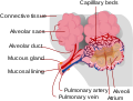 Alveolus diagram


-
 Anatomy of lungs.
 -
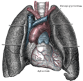 Front view of heart and lungs.
 -
 Transverse section of thorax, showing relations of pulmonary artery.


-
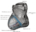 Base and diaphragmatic surface of heart.
 -
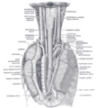 The position and relation of the esophagus in the cervical region and in the posterior mediastinum. Seen from behind.Crystal.
 -
 Pulmonary artery
 -
 Pulmonary trunk
 -
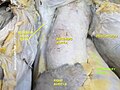
 Pulmonary artery
 -
 Pulmonary artery
 -
 Pulmonary trunk
 -
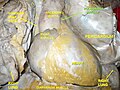
 Pulmonary artery
 -
 Pulmonary artery


See also
- Chronic obstructive lung disease
- Pulmonary hypertension
- Thromboembolic disease
- Pulmonary circulation
- Rasmussen's aneurysm
External links
- . GPnotebook https://www.gpnotebook.co.uk/simplepage.cfm?ID=53805116.
{{cite web}}: Missing or empty|title=(help) - Template:EMedicineDictionary
- Anatomy photo:20:01-0106 at the SUNY Downstate Medical Center - "Heart: The Pericardial sac and Great vessels"
- Anatomy photo:20:07-0105 at the SUNY Downstate Medical Center - "Heart: Openings of Great Vessels into the Pericardial Sac"
- Anatomy figure: 19:05-06 at Human Anatomy Online, SUNY Downstate Medical Center - "Mediastinal surface of the right lung."
- Anatomy figure: 19:06-02 at Human Anatomy Online, SUNY Downstate Medical Center - "Mediastinal surface of the left lung."
- Histology image: 13802loa – Histology Learning System at Boston University