
Structural coloration: Difference between revisions
m →Photonic crystals: chitin |
→Photonic crystals: (edited with ProveIt) |
||
| Line 59: | Line 59: | ||
==== Photonic crystals ==== |
==== Photonic crystals ==== |
||
[[Photonic crystal]]s can be formed in different ways.<ref>{{cite journal | url=http://perso.fundp.ac.be/~jpvigner/press/BioPhotonics/Beyond.pdf | title=Beyond butterflies—the diversity of biological photonic crystals | author=Welch, V.L., Vigneron, J.-P. | journal=Opt Quant Electron | year=2007 | month=July | volume=39 | pages=295-303 | doi=10.1007/s11082-007-9094-4}}</ref> |
|||
[[Photonic crystal]]s can be formed in different ways. |
|||
In ''Parides sesostris'', the Emerald-patched [[Cattleheart]] butterfly,<ref>{{cite web | url=http://optoelectronics.eecs.berkeley.edu/eliy_SCIAM.pdf | title=Photonic Crystals: Semiconductors of Light | publisher=Scientific American | date=December 2001 | accessdate=15 May 2012 | author=Yablonovitch, Eli | pages=46-55}}</ref> photonic crystals are formed of arrays of nano-sized holes in the chitin of the wing scales. The holes have a diameter of about 150 [[nanometre]]s and are about the same distance apart. The holes are arranged regularly in small patches; neighbouring patches contain arrays with differing orientations. The result is that these Emerald-patched Cattleheart scales reflect green light evenly at different angles instead of being iridescent.<ref name=Ball/><ref name="VukusicPhotonics">{{cite journal | title=Natural Photonics | author=Vukusic, P. | journal=Physics World | year=2004 | month=February | volume=17 | issue=2 | pages=35–39}}</ref> |
In ''Parides sesostris'', the Emerald-patched [[Cattleheart]] butterfly,<ref>{{cite web | url=http://optoelectronics.eecs.berkeley.edu/eliy_SCIAM.pdf | title=Photonic Crystals: Semiconductors of Light | publisher=Scientific American | date=December 2001 | accessdate=15 May 2012 | author=Yablonovitch, Eli | pages=46-55}}</ref> photonic crystals are formed of arrays of nano-sized holes in the chitin of the wing scales. The holes have a diameter of about 150 [[nanometre]]s and are about the same distance apart. The holes are arranged regularly in small patches; neighbouring patches contain arrays with differing orientations. The result is that these Emerald-patched Cattleheart scales reflect green light evenly at different angles instead of being iridescent.<ref name=Ball/><ref name="VukusicPhotonics">{{cite journal | title=Natural Photonics | author=Vukusic, P. | journal=Physics World | year=2004 | month=February | volume=17 | issue=2 | pages=35–39}}</ref> |
||
Revision as of 14:44, 19 May 2012

Structural coloration is the production of colour by microscopically structured surfaces fine enough to interfere with visible light, sometimes in combination with pigments: for example, peacock tail feathers are pigmented brown, but their structure makes them appear blue, turquoise, and green, and often they appear iridescent.[1][2] In animals, these colours are created in a range of ways, including by diffraction gratings and other photonic effects.
Structural coloration was first observed by English scientists Robert Hooke and Isaac Newton, and its principle – wave interference – explained by Thomas Young a century later.
History

Hooke
In his 1665 book Micrographia, Robert Hooke described the "fantastical" (structural, not pigment) colours of the Peacock's feathers:[1]
- "The parts of the Feathers of this glorious Bird appear, through the Microscope, no less gaudy then do the whole Feathers; for, as to the naked eye 'tis evident that the stem or quill of each Feather in the tail sends out multitudes of Lateral branches, ... so each of those threads in the Microscope appears a large long body, consisting of a multitude of bright reflecting parts.
... their upper sides seem to me to consist of a multitude of thin plated bodies, which are exceeding thin, and lie very close together, and thereby, like mother of Pearl shells, do not onely reflect a very brisk light, but tinge that light in a most curious manner; and by means of various positions, in respect of the light, they reflect back now one colour, and then another, and those most vividly. Now, that these colours are onely fantastical ones, that is, such as arise immediately from the refractions of the light, I found by this, that water wetting these colour'd parts, destroy'd their colours, which seem'd to proceed from the alteration of the reflection and refraction."
Newton
In his 1704 book Opticks, Isaac Newton described the mechanism of the colours (other than the brown pigment) of peacock tail feathers.[3] Newton noted that[4]
- "The finely colour'd Feathers of some Birds, and particularly those of Peacocks Tails, do, in the very same part of the Feather, appear of several Colours in several Positions of the Eye, after the very same manner that thin Plates were found to do in the 7th and 19th Observations, and therefore their Colours arise from the thinness of the transparent parts of the Feathers; that is, from the slenderness of the very fine Hairs, or Capillamenta, which grow out of the sides of the grosser lateral Branches or Fibres of those Feathers."
Young
Thomas Young (1773–1829) extended Newton's particle theory of light by showing that light could also behave as a wave. He showed in 1803 that light could diffract from sharp edges or slits, creating interference patterns.[5][6]
Beddard
.JPG)
In his 1892 book Animal Coloration, An Account of the Principal Facts and Theories Relating to the Colours and Marking of Animals, Frank Evers Beddard (1858–1925) acknowledged the existence of structural colours:
- "The colours of animals are due either solely to the presence of definite pigments in the skin, or ... beneath the skin; or they are partly caused by optical effects due to the scattering, diffraction or unequal refraction of the light rays. Colours of the latter kind are often spoken of as structural colours; they are caused by the structure of the coloured surfaces. The metallic lustre of the feathers of many birds, such as the humming birds, is due to the presence of excessively fine striae upon the surface of the feathers."[7]: 1
But Beddard then largely dismissed structural coloration, firstly as subservient to pigments: "in every case the [structural] colour needs for its display a background of dark pigment;"[7]: 2 and then by asserting its rarity: "By far the commonest source of colour in invertebrate animals is the presence in the skin of definite pigments ..."[7]: 2 , though he does later admit that the Cape Golden Mole has "structural peculiarities" in its hair that "give rise to brilliant colours".[7]: 32
Principles
Structure not pigment
Structural coloration is caused by interference effects rather than by pigments. Colours are produced when a material is scored with fine parallel lines, formed of one or more parallel thin layers, or otherwise composed of microstructures on the scale of the colour's wavelength. Structural coloration is responsible for the blues and greens of the feathers of many birds (the bee-eater, kingfisher and roller, for example), as well as many butterfly wings and beetle wing-cases (elytra).[2] These are often iridescent, as in peacock feathers and mother of pearl,[2] because the reflected colour depends on the viewing angle, which in turn governs the apparent spacing of the structures responsible.[8] Structural colours can be combined with pigment colours: peacock feathers are pigmented brown with melanin.
Principle of iridescence
Iridescence, as explained by Thomas Young in 1803, is created when extremely thin films reflect part of the light falling on them from their top surfaces. The rest of the light goes through the films, and a further part of it is reflected from their bottom surfaces. The two sets of reflected waves travel back upwards in the same direction. But since the bottom-reflected waves travelled a little further – controlled by the thickness and refractive index of the film, and the angle at which the light fell – the two sets of waves are out of phase. When the waves are one or more whole wavelength apart – in other words at certain specific angles, they add (interfere constructively), giving a strong reflection. At other angles and phase differences, they can subtract, giving weak reflections. The thin film therefore selectively reflects just one wavelength – a pure colour – at any given angle, but other wavelengths – different colours – at different angles. So, as a thin-film structure like a butterfly's wing or bird's feather moves, it seems to change colour.[3]
Structures can be far more elaborate than a single thin film: films can be stacked up to give strong iridescence, to combine two colours, or to balance out the inevitable change of colour with angle to give a more diffuse, less iridescent effect. Different methods by which animals produce structural colour are described below.[2]
Mechanisms
Fixed structures

Diffraction gratings
A diffraction grating constructed of layers of chitin and air gives rise to the iridescent colours of various butterfly wing scales as well as to the tail feathers of birds such as the peacock. Hooke and Newton were correct in their claim that the peacock's colours are created by interference, but the structures responsible, being close to the wavelength of light in scale (see micrographs), were smaller than the striated structures they could see with their light microscopes.[2]
Another way to produce a diffraction grating is with tree-shaped arrays of chitin, as in the wing scales of some of the brilliantly coloured tropical Morpho butterflies (see drawing).[2]
Yet another variant exists in Parotia lawesii, Lawes's Parotia, a bird of paradise. The barbules of the feathers of its brightly coloured breast patch are V-shaped, creating thin-film microstructures that strongly reflect two different colours, bright blue-green and orange-yellow. When the bird moves the colour switches sharply between these two colours, rather than drifting iridescently. During courtship, the male bird systematically makes small movements to attract females, so the structures must have evolved through sexual selection.[9][2]
Selective mirrors
Selective mirrors to create interference effects are formed of micron-sized bowl-shaped pits lined with multiple layers of chitin in the wing scales of Papilio palinurus, the Emerald Swallowtail butterfly. These act as highly selective mirrors for two wavelengths of light. Yellow light is reflected directly from the centres of the pits; blue light is reflected twice by the sides of the pits. The combination appears green, but can be seen as an array of yellow spots surrounded by blue circles under a microscope.[2]
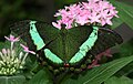
Photonic crystals
Photonic crystals can be formed in different ways.[10]
In Parides sesostris, the Emerald-patched Cattleheart butterfly,[11] photonic crystals are formed of arrays of nano-sized holes in the chitin of the wing scales. The holes have a diameter of about 150 nanometres and are about the same distance apart. The holes are arranged regularly in small patches; neighbouring patches contain arrays with differing orientations. The result is that these Emerald-patched Cattleheart scales reflect green light evenly at different angles instead of being iridescent.[2][12]
In Lamprocyphus augustus, a weevil from Brazil, the chitin exoskeleton is covered in iridescent green oval scales. These contain diamond-based crystal lattices oriented in all directions to give a brilliant green coloration that hardly varies with angle. The scales are effectively divided into pixels about a μmetre wide. Each such pixel is a single crystal and reflects light in a direction different from its neighbours.[13][14]
Crystal fibres
Crystal fibres formed of hexagonal arrays of hollow nanofibres are used to create the bright iridescent colours of the bristles (setae) Aphrodita, the Sea Mouse, a non-wormlike genus of marine annelids.[2] The colours are aposematic, warning predators not to attack.[15] The chitin walls of the hollow bristles form a hexagonal honeycomb-shaped photonic crystal; the hexagonal holes are 0.51 μmetre apart. The structure behaves optically as if it consisted of a stack of 88 diffraction gratings, making Aphrodita one of the most iridescent of marine organisms.[16]
Deformed matrices
Deformed matrices, consisting of randomly-oriented nanochannels in a spongelike keratin matrix, create the diffuse non-iridescent blue colour of Ara ararauna, the Blue-and-yellow Macaw. Since the reflections are not all arranged in the same direction, the colours, while still magnificent, do not vary much with angle, so they are not iridescent.[2][17]
Variable structures
Reversible proteins
Some animals including cephalopods like squid are able to vary their colours rapidly for both camouflage and signalling. The mechanisms include reversible proteins which can be switched between two configurations. The configuration of reflectin proteins in chromatophore cells in the skin of the Loligo pealeii squid is controlled by electric charge. When charge is absent, the proteins stack together tightly, forming a thin, more reflective layer; when charge is present, the molecules stack more loosely, forming a thicker layer. Since chromatophores contain multiple reflectin layers, the switch changes the layer spacing and hence the colour of light that is reflected.[2]
-
 In Morpho butterflies such as Morpho helena the brilliant colours are produced by intricate firtree-shaped microstructures too small for optical microscopes.
 -
 Ink drawing of the 'Christmas tree' thin layer optical interference structures in a Morpho butterfly wing scale. Each subtree has about 10 thin films, seen in cross section. From an electron micrograph.
 -
 The male Parotia lawesii bird of paradise signals to the female with his breast feathers that switch from blue to yellow.
 -
 Brilliant green of Emerald Swallowtail Papilio palinurus is created by arrays of microscopic bowls that reflect yellow directly and blue from the sides.
 -
 Emerald-patched cattleheart butterfly, Parides sesostris, creates its brilliant green using photonic crystals.
 -
 Iridescent scales of Lamprocyphus augustus weevil contain diamond-based crystal lattices oriented in all directions to give almost uniform green.
 -
 Hollow nanofibre bristles of Aphrodita aculeata (a species of Sea mouse) reflect light in yellows, reds and greens to warn off predators.
 -
 Magnificent non-iridescent colours of Blue-and-yellow Macaw are created by random nanochannels.
 -
 Longfin Inshore Squid, Loligo pealeii has been studied for its ability to change colour.
_KL.jpg)


_-_Relic38.jpg)


.jpg)


In technology
Structural coloration could be exploited industrially and commercially, and research that could lead to such applications is under way.[2] A direct parallel would be to create military camouflage fabrics that vary their colours and patterns to match their environments, just as chameleons and cephalopods do.[2] The ability to vary reflectivity to different wavelengths of light could also lead to efficient optical switches that could function like transistors, enabling engineers to make fast optical computers and routers.[2]
See also
Bibliography
Pioneering books
- Beddard, F.E. Animal Coloration, An Account of the Principal Facts and Theories Relating to the Colours and Markings of Animals. Swan Sonnenschein, London, 1892. 2nd Edition, 1895.
- Hooke, R. Micrographia, John Martyn and James Allestry, London, 1665.
- Newton, I. Opticks, William Innys, London, 1704.
Research
- Fox, D.L. Animal Biochromes and Animal Structural Colours. University of California Press, 1992.
- Johnsen, S. The Optics of Life: A Biologist's Guide to Light in Nature. Princeton University Press, 2011.
- Kolle, M. Photonic Structures Inspired by Nature . Springer, 2011.
General books
- Brebbia, C.A. Colour in Art, Design and Nature. WIT Press, 2011.
- Lee, D.W. Nature's Palette: The Science of Plant Color. University of Chicago Press, 2008.
References
- ^ a b Hooke, R. Micrographia. Chapter 36 ('Observ. XXXVI. Of Peacoks, Ducks, and Other Feathers of Changeable Colours.')
- ^ a b c d e f g h i j k l m n o Ball, Philip (May 2012). "Scientific American". Nature's Color Tricks. Vol. 306. pp. 74–79. doi:10.1038/scientificamerican0512-74. Retrieved April 23, 2012.
- ^ a b "Iridescence in Lepidoptera". Photonics in Nature (originally in Physics Review). University of Exeter. September 1998. Retrieved April 27, 2012.
- ^ Newton, Isaac (Fourth Edition, 1730. (First published 1704)). "Opticks". William Innys at the West-End of St. Paul's, London. pp. Prop. V., page 251. Retrieved April 27, 2012.
{{cite web}}: Check date values in:|date=(help) - ^ Young, Thomas (1804). "Experimental Demonstration of the General Law of the Interference of Light". Philosophical Transactions of the Royal Society of London. 94.
- ^ Shamos, Morris (1959). Great Experiments in Physics. New York: Holt Reinhart and Winston. pp. 96–101.
- ^ a b c d Frank Evers Beddard. Animal Coloration. 1892.
- ^ Wallin, Margareta (2002). "Nature's Palette" (PDF). Nature's Palette: How animals, including humans, produce colours. Bioscience-explained.org. pp. Vol 1, No 2, pages 1–12. Retrieved November 17, 2011.
- ^ Stavenga, Doekele G. (2010). "Dramatic colour changes in a bird of paradise caused by uniquely structured breast feather barbules". Proceedings of the Royal Society B. 278: 2098–2104. doi:10.1098/rspb.2010.2293.
- ^ Welch, V.L., Vigneron, J.-P. (2007). "Beyond butterflies—the diversity of biological photonic crystals" (PDF). Opt Quant Electron. 39: 295–303. doi:10.1007/s11082-007-9094-4.
{{cite journal}}: Unknown parameter|month=ignored (help)CS1 maint: multiple names: authors list (link) - ^ Yablonovitch, Eli (December 2001). "Photonic Crystals: Semiconductors of Light" (PDF). Scientific American. pp. 46–55. Retrieved 15 May 2012.
- ^ Vukusic, P. (2004). "Natural Photonics". Physics World. 17 (2): 35–39.
{{cite journal}}: Unknown parameter|month=ignored (help) - ^ Galusha, Jeremy W., Lauren R. Richey, John S. Gardner, Jennifer N. Cha, Michael H. Bart (2008). "Discovery of a diamond-based photonic crystal structure in beetle scales". Physical Review E. 77 (5): 050904. doi:10.1103/PhysRevE.77.050904.
{{cite journal}}: Unknown parameter|month=ignored (help)CS1 maint: multiple names: authors list (link) - ^ Biomimicry News: The photonic Beetle. 21 May 2008
- ^ "Sea mouse promises bright future". BBC News. BBC. January 3, 2001. Retrieved April 26, 2012.
- ^ McPhedran, Ross; McKenzie, David; Nicorovici, Nicolae (3 April 2002). "A Natural Photonic Crystal" (PDF). University of Sydney School of Physics. Retrieved May 18, 2012.
{{cite web}}: CS1 maint: multiple names: authors list (link) Contains good micrographs. - ^ Vukusic, P., Sambles, J.R. (2003). "Photonic Structures in Biology". Nature. 424: 852–855.
{{cite journal}}: Unknown parameter|month=ignored (help)CS1 maint: multiple names: authors list (link)
.jpg)