Amoeba (genus)
| Amoeba | |
|---|---|

| |
| Scientific classification | |
| Domain: | |
| Kingdom: | |
| Phylum: | |
| Order: | |
| Family: | |
| Genus: | Amoeba Bory de Saint-Vincent, 1822
|
| Species | |
Amoeba (sometimes amœba or ameba, plural amoebae or amoebas) is a genus of Protozoa.[1] It is a shapeless unicellular organism.
History
The amoeba was first discovered by August Johann Rösel von Rosenhof in 1757.[2] Early naturalists referred to Amoeba as the Proteus animalcule after the Greek god Proteus, who could change his shape. The name "amibe" was given to it by Bory de Saint-Vincent,[3] from the Greek amoibè (αμοιβή), meaning change.[4] Dientamoeba fragili was first described in 1918, and was linked to harm in humans.[5]
Anatomy

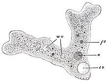
The cell's organelles and cytoplasm are enclosed by a cell membrane; it obtains its food through phagocytosis. This makes amoebae heterotrophs. Amoebae have a single large tubular pseudopod at the anterior end, and several secondary ones branching to the sides. The most famous species, Amoeba proteus, averages about 220-740 μm in length, while in motion.Cite error: The <ref> tag has too many names (see the help page). making it a giant among amoeboids.[6] A few amoeboids belonging to different genera can grow larger, however, such as Gromia, Pelomyxa, and Chaos.
Amoebae's most recognizable features include one or more nuclei and a simple contractile vacuole to maintain osmotic equilibrium. Food enveloped by the amoeba is stored and digested in vacuoles. Amoebae, like other unicellular eukaryotic organisms, reproduce asexually via mitosis and cytokinesis, not to be confused with binary fission, which is how prokaryotes (bacteria) reproduce. In cases where the amoeba are forcibly divided, the portion that retains the nucleus will survive and form a new cell and cytoplasm, while the other portion dies. Amoebae also have no definite shape.[7]
Genome
The amoeba is remarkable for its very large genome. The species Amoeba proteus has 290 billion base pairs in its genome, while the related Polychaos dubium (formerly known as Amoeba dubia) has 670 billion base pairs. The human genome is small by contrast, with its count of 2.9 billion base pairs.[8]
Osmoregulation
Like most other protists, amoebas have a contractile vacuole complex. Amoeba proteus, a free-living, freshwater species of amoeba, has one contractile vacuole (CV), which is a membrane-bound organelle. The CV slowly fills with water from the cytoplasm (diastole), and, while fusing with the cell membrane, it quickly contracts releasing water to the outside (systole) by exocytosis. This process regulates the amount of water present in the cytoplasm of the amoeba; it is, therefore, a means of osmoregulation.
Immediately after the CV expels water, its membrane crumples, and soon afterwards, many small vacuoles or vesicles appear surrounding the membrane of the CV.[9] It is suggested that these vesicles split from the CV membrane itself. The small vesicles gradually increase in size as they take in water and then they fuse with the CV, which grows in size as it fills with water. Therefore, the function of these numerous small vesicles is to collect excess cytoplasmic water and channel it to the central CV. The CV swells for a number of minutes and then contracts to expel the water outside. The cycle is then repeated again
The membranes of the small vesicles as well as the membrane of the CV have aquaporin proteins embedded in them.[9] These transmembrane proteins facilitate water passage through the membranes. The presence of aquaporin proteins in both CV and the small vesicles suggests that water collection occurs both through the CV membrane itself as well as through the function of the vesicles. However, the vesicles, being more numerous and smaller, would allow a faster water uptake due to the larger total surface area provided by the vesicles.[9]
The small vesicles also have another protein embedded in its membrane: Vacuolar-type H+-ATPase or V-ATPase.[9] This ATPase pumps H+ ions into the vesicle lumen, lowering its pH with respect to the cytosol. However, the pH of the CV in some amoebas is only mildly acidic, suggesting that the H+ ions are being removed from the CV or from the vesicles. It is thought that the electrochemical gradient generated by V-ATPase might be used for the transport of ions (it is presumed K+ and Cl-) into the vesicles. This builds an osmotic gradient across the vesicle membrane, leading to influx of water from the cytosol into the vesicles by osmosis,[9] which is facilitated by aquaporins.
Since these vesicles fuse with the central contractile vacuole, which expels the water, ions end up being removed from the cell, which is not beneficial for a freshwater organism. The removal of ions with the water has to be compensated by some yet-unidentified mechanism.
Like most cells, amoebae are adversely affected by excessive osmotic pressure caused by extremely saline or dilute water. Amoebae will prevent the influx of salt in saline water, resulting in a net loss of water as the cell becomes isotonic with the environment, causing the cell to shrink. Placed into fresh water, amoebae will also attempt to match the concentration of the surrounding water, causing the cell to swell and sometimes burst if the water surrounding the amoeba is too dilute.[10]
Amoebic cysts
In environments that are potentially lethal to the cell, an amoeba may become dormant by forming itself into a ball and secreting a protective membrane to become a microbial cyst. The cell remains in this state until it encounters more favourable conditions.[7] While in cyst form the amoeba will not replicate and may die if unable to emerge for a lengthy period of time.
Nutrition in amoeba
Amoeba obtains its nutrition in a heterotrophic mode. Both the anabolic and catabolic functions are carried out in the same cell. Amoeba feeds on plankton and diatoms present in water. It can form arm- like structures called pseudopodia, extending from any part of its body as it is shapeless. When it senses food in its surroundings it extends its pseudopodia in that direction and moves towards it. Then it engulfs the food with its pseudopodia. When the food enters its body the amoeba forms a food vacuole around it which contains certain enzymes to digest the food. When the food is digested the unwanted waste is released through its body surface.
Video gallery
References
- ^ "Amoeba" at Dorland's Medical Dictionary
- ^ Leidy, Joseph (1878). "Amoeba proteus". The American Naturalist. 12 (4): 235–238. doi:10.1086/272082. JSTOR 2463786.
- ^ Audouin, Jean-Victor (1826). Dictionnaire classique d'histoire naturelle. Rey et Gravier. p. 5.
{{cite book}}: Unknown parameter|coauthors=ignored (|author=suggested) (help) - ^ McGrath, Kimberley (2001). Gale Encyclopedia of Science Vol. 1: Aardvark-Catalyst (2nd ed.). Gale Group. ISBN 0-7876-4370-X. OCLC 46337140.
{{cite book}}: Unknown parameter|coauthors=ignored (|author=suggested) (help) - ^ Eugene H. Johnson, Jeffrey J. Windsor, and C. Graham Clark Emerging from Obscurity: Biological, Clinical, and Diagnostic Aspects of Dientamoeba fragilis.
- ^ MacIver, Sutherland. "Isolation of Amoebae". The Amoebae. Retrieved 2009-10-08.
- ^ a b "Amoeba". Scienceclarified.com.
- ^ http://www.genomenewsnetwork.org/articles/02_01/Sizing_genomes.shtml
- ^ a b c d e Nishihara, E., Yokota, E., Tazaki, A., Orii, H., Katsuhara, M., Kataoka, K., et al. (2008). Presece of aquaporin and V-ATPase on the contractile vacuole of Amoeba proteus. Biology of the cell , 100 (3), 179-188.
- ^ Patterson, D.J. (1981). "Contractile vacuole complex behaviour as a diagnostic character for free living amoebae". Protistologica. 17: 243–248.
