
Human pharynx: Difference between revisions
| Line 18: | Line 18: | ||
One current scheme places the following traditional phyla in [[Platyzoa]]: [[Platyhelminthes]], [[Gastrotricha]], [[Gnathifera]], [[Rotifera]], [[Acanthocephala]], [[Gnathostomulida]], [[Micrognathozoa]], and [[Cycliophora]]. The pharyngeal system within this superphyllum can include a cilial lining, the ability to [[Eversion|evert]] the pharynx, secretion, powerful muscles, a jaw-like structure, bilateral symmetry, and lateral lamellae. |
One current scheme places the following traditional phyla in [[Platyzoa]]: [[Platyhelminthes]], [[Gastrotricha]], [[Gnathifera]], [[Rotifera]], [[Acanthocephala]], [[Gnathostomulida]], [[Micrognathozoa]], and [[Cycliophora]]. The pharyngeal system within this superphyllum can include a cilial lining, the ability to [[Eversion|evert]] the pharynx, secretion, powerful muscles, a jaw-like structure, bilateral symmetry, and lateral lamellae. |
||
==''Gastrotricha'' myoepithelial pharynx== |
|||
The myoepithelial pharynx of the Gastrotricha is an important distinguishing feature.<ref name=Hochberg>{{cite journal |author=Hochberg R, Litvaitis MK |title=Phylogeny of Gastrotricha: a morphology-based framework of gastrotrich relationships |journal=Biol Bull. |volume=198 |issue=2 |pages=299-304 |year=2000 |month=Apr |pmid= 10786949 }}</ref> In the order ''Macrodasyida'' the pharynx has pharyngeal pores, while in the other order ''Chaetonotida'', there is a pharyngeal plug.<ref name=Hochberg/> The myoepithelial pharynx is also triradiate.<ref name=Hochberg/> |
|||
*[http://www.biolbull.org/cgi/reprint/198/2/299 Schematic representations of the two orders within Gastrotricha showing the difference in pharynges.] |
|||
==''Platyhelminthes'' pharynx== |
==''Platyhelminthes'' pharynx== |
||
Revision as of 05:45, 13 February 2009
The pharynx (plural: pharynges) is part of the neck and throat situated immediately posterior to (behind) the mouth and nasal cavity, and cranial, or superior, to the oesophagus (esophagus), larynx, and trachea.
Pharyngeal systems
In anatomy and zoology the pharynx is that part of the alimentary canal immediately behind the mouth. It is a membrane-lined cavity also behind the nose (when present) which connects to the esophagus.
A pharyngeal system may contain tissues and organs that are not found in the pharynx of an adult human. These tissues and organs may be considered as part of the pharynx due to anatomical or physiological inferences or deductions. As additional analytical techniques become available, they may be able to indicate whether those inferences or deductions are incorrect, perhaps incomplete, or correct. Phylogenetic analysis using molecular genetics and genomics is one such technique.
Gene expression in eukaryotes can often help to establish that particular organs occur at some developmental stage. A mature mRNA template is one occurring in a cell where a gene is expressed as part of a particular cell's function. Reverse transcriptase scans the mature mRNA and synthesizes a sequence of DNA that complements the mRNA template. This strand of DNA is complementary DNA (cDNA). Finding cDNA in Unigene, for example
- [1],
can help to establish that specific genes are expressed in tissues or organs such as the pharynx. Expression of these same or similar genes in organisms of questionable classification can help in their taxonomy.
Platyzoa pharyngeal system
One current scheme places the following traditional phyla in Platyzoa: Platyhelminthes, Gastrotricha, Gnathifera, Rotifera, Acanthocephala, Gnathostomulida, Micrognathozoa, and Cycliophora. The pharyngeal system within this superphyllum can include a cilial lining, the ability to evert the pharynx, secretion, powerful muscles, a jaw-like structure, bilateral symmetry, and lateral lamellae.
Gastrotricha myoepithelial pharynx
The myoepithelial pharynx of the Gastrotricha is an important distinguishing feature.[1] In the order Macrodasyida the pharynx has pharyngeal pores, while in the other order Chaetonotida, there is a pharyngeal plug.[1] The myoepithelial pharynx is also triradiate.[1]
- Schematic representations of the two orders within Gastrotricha showing the difference in pharynges.
Platyhelminthes pharynx
Some of the turbellarians have a simple pharynx lined with cilia. Others can evert their pharynx to envelope prey. Some species of platyhelminths break up and soften food first by secreting enzymes in the gut or the pharynx (throat).[2]
Adult Digenea use a relatively large, muscular pharynx to ingest cells, cell fragments, mucus, body fluids or blood.
In some species of Monogenea the pharynx secretes enzymes that digest the host's skin, allowing the parasite to feed on blood and cellular debris.
Rotifera mastax
Rotifaers have several ciliated tufts around the mouth that in motion resemble a wheel. These create a current that sweeps food into the mouth, where it is chewed up by a characteristic pharynx (called the mastax) containing a tiny, calcified, jaw-like structure called the trophi[3][4].
Acanthocephala
Acanthocephalans are highly specialized parasites without mouth, pharynx and gut. Phylogenetic analysis of the 18S ribosomal gene has revealed that the Acanthocephala are most closely related to the rotifers, or may even belong in that phylum.

Gnathostomulida pharynx
The gnathostomulids are characterized by a specialized, muscular jaw, which they use to scrape smaller organisms off of the grains of sand that make up their anoxic seabed mud habitat.[5] This bilaterally symmetrical pharynx with its complex cuticular mouth parts make them appear closely related to rotifers and their allies, together making up the Gnathifera.
Micrognathozoa pharyngeal apparatus
Micrognathozoa show strong affinities with both Rotifera and Gnathostomulida (within the taxon Gnathifera), especially in the fine structure of the pharyngeal apparatus, where the jaw elements have cuticular rods with osmiophilic cores.[6] In Limnognathia maerski the main parts of the complicated jaw system have two lateral pharyngeal lamellae.[6]
Lophotrochozoa pharynx
The Lophophorata include the Bryozoa, Entoprocta, Phoronida, and Brachiopoda.
The lophophorates all feed by "impingement".[7]
The Trochozoa include the Nemertea, Mollusca, Sipuncula, Echiura, and Annelida.
Bryozoa pharynx
The Bryozoan gut is U-shaped, and consists of a pharynx which passes into the esophagus and cardia. The mouth opens into a relatively short, large, and muscular pharynx with a thick wall of tall columnar epithelial cells. The anterior region of the pharynx, into which the mouth opens, is ciliated. Food particles accumulate in the pharynx before being passed through the esophagus and cardia to the stomach.
In Gymnolaemata the feeding current is directed by the lophophore at the mouth where particles impinge and are sucked into the pharynx.[7] In Phylactolaemata particles impinge in the trough of the lophophore and are transported to the pharynx by cilia.[7]
Phoronida pharynx
In the Phoronids particles impinge on the frontal surface of the tentacles and are conveyed to the pharynx by tracts of cilia.[7]

Brachiopoda pharynx
In Brachiopods particles impinge on the frontal surface of the tentacles and are conveyed to the pharynx by tracts of cilia.[7]
Nemertea pharynx
The pharynx in the Nemertea genus Cerebratulus has an ectodermal pharynx that is the first portion of the gut.[8]
Mollusca pharynx
Mollusks have pharyngeal glands (sugar glands) situated laterally to the pharynx.[9]
Annelida pharynx
The Annelids, for example earthworms, have a pharynx. It is relatively strong due to the fact that the wall of the pharynx consists of epithelium with built up obliquely striated longitudinal and circular muscle layers between it and the nervous tissue. Earthworms, like many other annelids, suck their food. This sucking action is aided by the pharyngeal muscles.

In leeches, like the earthworm, the pharynx extends from the mouth to the esophagus which connects to the crop.
Ecdysozoa triradiate pharynx
The Ecdysozoa include the following phyla: Arthropoda, Onychophora, Tardigrada, Kinorhyncha, Priapulida, Loricifera, Nematoda and Nematomorpha.
Ecdysozoans have various features are found in the group, for instance, tardigrades, pycnogonids and roundworms have a triradiate pharynx.
Priapulida everted pharynx
The mouth cone (“everted pharynx”) of a possible new species of Meiopriapulus bears pharyngeal teeth.[10]
Loricifera pharyngeal apparatus
Loriciferans have unique adult features including the cuticular buccal tube and myoepithelial pharyngeal apparatus with placoids.[6] Annulation of the flexible buccal tube, telescoping of the mouth cone and three rows of placoids in the triradiated pharynx bulb are found only in Tardigrada and Loricifera.[6] The phylum Loricifera has several plesiomorphic characters, such as a myoepithelial pharyngeal bulb, comparable to the other Cycloneuralia, especially the Nematoda and Gastrotricha.[6] The parasitic Nematomorpha, whose adults lack the pharyngeal bulb, has a larva with an introvert similar to Scalidophora. The loriciferans have, therefore, been assigned to the Cephalorhyncha that includes Priapulida, Kinorhyncha and Nematomorpha.[6]
Tardigrada pharynx bulb
The pharynx is of a triradiate, muscular, sucking kind, armed with stylets. Annulation of the flexible buccal tube, telescoping of the mouth cone and three rows of placoids in the triradiated pharynx bulb are found only in Tardigrada and Loricifera.[6]
The Ecdysozoa-concept resolves the problem of the nematode-like pharynx and some data from 18S-rRNA and the HOX (homeobox) gene indicate a relation to roundworms.
Echinodermata pharyngeal systems
All five chordate characteristics are lacking in echinoderms.[11] Echinodermata have not as yet been reported to express the transcription factor Pax1/9.[11]
Ophiuroidea (brittle stars and basket stars)
Asteroidea (sea stars)
Echinoidea (sea urchins) pharynx
In images of the sea urchin (Strongylocentrous purpuratus) larva, produced with differential interference contrast optics, the pharynx is clearly visible and labelled.[12]
The pharynx in the sea urchin genuses Evechinus and Echinus is part of the alimentary canal, just after the mouth, and enclosed within Aristotle's lantern.[13] The lantern must be opened by dissection to observe the pharynx.[13] The pharynx leads into the narrow, much convoluted oesophagus.[13] The external covering of the pharynx is ciliated epithelium.[13]
Holothuroidea (sea cucumbers) pharyngeal bulb
In sea cucumbers an important feature is a calcareous ring that encircles the pharynx or throat.[14] The muscles that operate the oral tentacles are attached to this ring.[14]
- The main internal anatomical features of the sea cucumber Dendrochirotida include a pharyngeal bulb.
Chordata pharyngeal system
The chordates have evolved a unique body plan within the deuterostomes and are considered to share five morphological characters: (1) a muscular postanal tail, (2) a notochord, (3) a dorsal neural tube, (4) an endostyle, and (5) pharyngeal gill slits.[11] Pharyngeal gills appear to function for feeding and oxygen absorption.[11] With the exception of tunicates, all deuterostomes with gills have structural elements associated with pharyngeal gills.[11] Studies of phylogeny have shown that it is likely the ancestral deuterostome was a worm that had pharyngeal gills.[11]
Xenoturbellida pharyngeal elements
All five chordate characteristics (postanal tail, dorsal nerve cord, notochord, endostyle, and pharyngeal gill slits) are lacking in Xenoturbella.[11] Xenoturbella have not been reported to express the transcription factor Pax1/9.[11]
Hemichordata pharyngeal elements
Three of the chordate characteristics are present in hemichordates: a ventral postanal tail, an endostyle, and pharyngeal gill slits.[11] Adult pharyngeal clefts have allied hemichordates with chordates.[15]
The hemichordate epibranchial ridge, like the chordate endostyle, is composed of specialized secretory cells, and these cells bind iodine. However, iodine binding occurs all throughout the pharynx.[11]
The hemichordate homolog of the gene NK2.1, which in chordates is expressed in the endostyle and the dorsal nervous system, is expressed in hemichordate larval neuronal structures and broadly in the pharyngeal endoderm, stomochord, and hindgut.[11] It appears that the endostyle function in chordates is accomplished broadly by the pharynx in hemichordates.[11]
The transcription factor Pax1/9 is expressed in the pharyngeal endoderm in hemichordates, cephalochordates, and in the vertebrate gnathostomes.[11]
Pharyngeal skeletal elements of hemichordates and cephalochordates are strikingly similar in appearance.[11]
Urochordata pharyngeal elements
Pharyngeal gill slits are present in tunicates only in the tunicate tadpole larva.[11] The endostyle is an iodine-binding organ present in the tunicate pharynx.[11]
Cephalochordata pharyngeal elements

Cephalochordates possess a pharynx.[11] The endostyle as an iodine-binding organ is also present in the cephalochordate pharynx.[11]
Vertebrata pharyngeal system
The expression of the most anterior Hox gene, Hox1, is first seen in vertebrates in the second pharyngeal slit and the expression of Hox1 in hemichordates is seen at the level between the first and second gill slits.[11]
Neural crest cartilages are found in the vertebrate pharyngeal gills, and neural crest cells do take on the role of pharyngeal cartilage secretion.[11] The pharyngeal gills are supported by collagenous cartilage skeletons in the hemichordates, cephalochordates, and vertebrates.[11]
Human pharynx
| Pharynx | |
|---|---|
 Head and neck. | |
 Pharynx | |
| Details | |
| Artery | pharyngeal branches of ascending pharyngeal artery, ascending palatine, descending palatine, pharyngeal branches of inferior thyroid |
| Vein | pharyngeal veins |
| Nerve | pharyngeal plexus |
| Identifiers | |
| Latin | Pharyngis |
| Anatomical terminology | |
Functions in humans
It is part of the digestive system and respiratory system of many organisms.
Because both food and air pass through the pharynx, a flap of connective tissue called the epiglottis closes over the trachea when food is swallowed to prevent choking or aspiration. In addition, some languages, such as Arabic, use the pharynx to articulate some sounds.
In persons with hayfever, oral allergy syndrome and related allergies, the pharynx is often a reaction site to allergens, with common symptoms including burning and itching.
Parts
The human pharynx is conventionally divided into three sections:
Oropharynx
The oropharynx lies behind the oral cavity and is lined by stratified squamous epithelium. The anterior wall consists of the base of the tongue and the vallecula; the lateral wall is made up of the tonsil, tonsillar fossa, and tonsillar (faucial) pillars; the superior wall consists of the inferior surface of the soft palate and the uvula.
Nasopharynx
The nasopharynx lies behind the nasal cavity.
Postero-superiorly this extends from the level of the junction of the hard and soft palates to the base of skull, laterally to include the fossa of Rosenmüller.
The inferior wall consists of the superior surface of the soft palate. Like the nasal passages it is lined with pseudostratified ciliated columnar epithelium.
Laryngopharynx
The laryngopharynx, also known as the hypopharynx, roughly corresponds to the levels between C4 to C6, it includes the pharyngo-esophageal junction (postcricoid area), the piriform sinus, and the posterior pharyngeal wall.
Like the oropharynx above it the hypopharynx serves as a passageway for food and air and is lined with a stratified squamous epithelium.
It lies inferior to the upright epiglottis and extends to the larynx, where the respiratory and digestive pathways diverge.
At that point, the laryngopharynx is continuous with the esophagus posteriorly. The esophagus conducts food and fluids to the stomach; air enters the larynx anteriorly. During swallowing, food has the "right of way", and air passage temporarily stops.
Inflammation
Pharyngitis is an inflammation of the throat or pharynx.[16] In most cases it is painful, and thus is often referred to as a sore throat. Inflammation of the tonsils (tonsillitis) and/or larynx (laryngitis) may occur simultaneously, which can make eating difficult or painful.
Most cases are caused by viral infections (40%-60%), with the remainder caused by bacterial infections, fungal infections, or irritants such as pollutants or chemical substances.[17]
Treatment of viral causes are mainly symptomatic while bacterial or fungal causes may be amenable to antibiotics and antifungals respectively.
Additional images
-
 Conducting passages.
 -
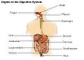 Organs of the digestive system.
 -
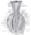
 The entrance to the larynx, viewed from behind.
 -
 Sagittal section of nose mouth, pharynx, and larynx.
 -
 The position and relation of the esophagus in the cervical region and in the posterior mediastinum. Seen from behind.


See also
References
- ^ a b c Hochberg R, Litvaitis MK (2000). "Phylogeny of Gastrotricha: a morphology-based framework of gastrotrich relationships". Biol Bull. 198 (2): 299–304. PMID 10786949.
{{cite journal}}: Unknown parameter|month=ignored (help) - ^ Walker, J.C., and Anderson, D.T. (2001). "The Platyhelminthes". In Anderson, D.T., (ed.). Invertebrate Zoology. Oxford University Press. pp. 58–80. ISBN 0195513681.
{{cite book}}: CS1 maint: extra punctuation (link) CS1 maint: multiple names: authors list (link) - ^ Klusemann J, Kleinow W, Peters W (1990). "The hard parts (trophi) of the rotifer mastax do contain chitin: evidence from studies on Brachionus plicatilis". Histochemistry. 94 (3): 277–83. PMID 2401635.
{{cite journal}}: Unknown parameter|month=ignored (help)CS1 maint: multiple names: authors list (link) - ^ Fontaneto D, Melone G (2006). "Postembryonic development of hard jaws (trophi) in a species belonging to the Brachionus plicatilis complex (Rotifera, Monogononta): a morphometric analysis". Microsc Res Tech. 69 (4): 296–301. PMID 16586490.
- ^ Barnes, R.F.K. et al (2001). The Invertebrates: A Synthesis. Oxford: Blackwell Science.
- ^ a b c d e f g Kristensen RM (2002). "An Introduction to Loricifera, Cycliophora, and Micrognathozoa". Integr Comp Biol. 42 (3): 641–51. doi:10.1093/icb/42.3.641.
- ^ a b c d e Bullivant JS (1968). "THE METHOD OF FEEDING OF LOPHOPHORATES (BRYOZOA, PHORONIDA, BRACHIOPODA)". NZ J Mar Freshwat Res. 2: 135–46.
{{cite journal}}: Cite has empty unknown parameter:|month=(help) - ^ "Cerebratulus Ribbonworm Invertebrate Anatomy OnLine".
- ^ Sturm, Charles F (2006). The Mollusks: A Guide to Their Study, Collection, and Preservation. Universal-Publishers. p. 220. ISBN 1581129300, 9781581129304.
{{cite book}}: Check|isbn=value: invalid character (help); More than one of|pages=and|page=specified (help); Unknown parameter|coauthors=ignored (|author=suggested) (help) - ^ Park JK, Rho HS, Kristensen RM, Kim W, Giribet G (2006). "First Molecular Data on the Phylum Loricifera – An Investigation into the Phylogeny of Ecdysozoa with Emphasis on the Positions of Loricifera and Priapulida". Zoolog Sci. 23 (11): 943–54. doi:10.2108/zsj.23.943. PMID 17189906.
{{cite journal}}: Unknown parameter|month=ignored (help)CS1 maint: multiple names: authors list (link) - ^ a b c d e f g h i j k l m n o p q r s t u Rychel AL, Smith SE, Shimamoto HT, Swalla BJ (2006). "Evolution and development of the chordates: collagen and pharyngeal cartilage". Mol Biol Evol. 23 (3): 541–9. doi:10.1093/molbev/msj055. PMID 16280542.
{{cite journal}}: Unknown parameter|month=ignored (help)CS1 maint: multiple names: authors list (link) - ^ Arenas-Mena C, Cameron AR, Davidson EH (2000). "Spatial expression of Hox cluster genes in the ontogeny of a sea urchin". Development. 127 (21): 4631–43. PMID 11023866.
{{cite journal}}: Unknown parameter|month=ignored (help)CS1 maint: multiple names: authors list (link) - ^ a b c d McRae A (1959). "Evechinus chloroticus (Val.), an Endemic New Zealand Echinoid". Trans Proc Roy Soc New Zealand 1868-1961. 86 (3 & 4): 205–67.
{{cite journal}}: Unknown parameter|month=ignored (help) - ^ a b "Holothuroidea Sea cucumbers".
- ^ Blair JE, Hedges SB (2005). "Molecular Phylogeny and Divergence Times of Deuterostome Animals". Mol Biol Evol. 22 (11): 2275–84. doi:10.1093/molbev/msi225. PMID 16049193.
{{cite journal}}: Unknown parameter|month=ignored (help) - ^ "pharyngitis" at Dorland's Medical Dictionary
- ^ "eMedicine - Pharyngitis : Article by John R Acerra".
- Template:Stedman's
- Human Anatomy and Physiology Elaine N. Marieb and Katja Hoehn, Seventh Edition.
- TNM Classification of Malignant Tumours Sobin LH & Wittekind Ch (eds)Sixth edition UICC 2002 ISBN 0-471-22288-7
External links
- Anatomy photo:31:st-1401 at the SUNY Downstate Medical Center