
Urethra: Difference between revisions
m r2.7.1) (Robot: Adding ro:Uretră |
Manglamchen (talk | contribs) |
||
| Line 98: | Line 98: | ||
==External links== |
==External links== |
||
* [http://reconstructiveurology.wordpress.com/ Reconstructive Urology] |
|||
* {{KansasHistology|epithel|epith07}} "Male Urethra" |
* {{KansasHistology|epithel|epith07}} "Male Urethra" |
||
<!-- |
|||
{{System and organs}} |
{{System and organs}} |
||
Revision as of 09:29, 10 February 2012
This article needs additional citations for verification. (June 2007) |
In anatomy, the urethra (from Greek οὐρήθρα - ourethra) is a tube that connects the urinary bladder to the genitals for the removal of fluids out of the body. In males, the urethra travels through the penis, and carries semen as well as urine. In females, the urethra is shorter and emerges above the vaginal opening.
The external urethral sphincter is a striated muscle that allows voluntary control over urination.
Anatomy
Female urethra
In the human female, the urethra is about 1.5–2 inches (4–5 cm) long and exits the body between the clitoris and the vagina, extending from the internal to the external urethral orifice. It is placed behind the symphysis pubis, embedded in the anterior wall of the vagina, and its direction is obliquely downward and forward; it is slightly curved with the concavity directed forward. Its lining is composed of stratified squamous epithelium, which becomes transitional near the bladder. The urethra consists of three coats: muscular, erectile, and mucous, the muscular layer being a continuation of that of the bladder. Between the superior and inferior fascia of the urogenital diaphragm, the female urethra is surrounded by the Sphincter urethrae (urethral sphincter). Somatic (conscious) innervation of the external urethral sphincter is supplied by the pudendal nerve. The uro-genital sinus may be divided into three component parts. The first of these is the cranial portion which is continuous with the allantois and forms the bladder proper. The pelvic part of the sinus forms the prostatic urethra and epithelium as well as the membranous urethra and bulbo urethral glands in the male and the membranous urethra and part of the vagina in females. The area above and on both sides of the female urethra is thought by some[who?] to be sexually sensitive and is sometimes referred to as the U-spot or urethral erogenous zone.
Male urethra
In the human male, the urethra is about 8 inches (20 cm) long and opens at the end of the penis. The urethra provides an exit for urine as well as semen during ejaculation.
The urethra is divided into four parts in men, named after the location:
| Region | Description | Epithelium |
| pre-prostatic urethra | This is the intramural part of the urethra and varies between 0.5 and 1.5 cm in length depending on the fullness of the bladder. | Transitional |
| prostatic urethra | Crosses through the prostate gland. There are several openings: (1) the ejaculatory duct receives sperm from the vas deferens and ejaculate fluid from the seminal vesicle, (2) several prostatic ducts where fluid from the prostate enters and contributes to the ejaculate, (3) the prostatic utricle, which is merely an indentation. These openings are collectively called the verumontanum. | Transitional |
| membranous urethra | A small (1 or 2 cm) portion passing through the external urethral sphincter. This is the narrowest part of the urethra. It is located in the deep perineal pouch. The bulbourethral glands (Cowper's gland) are found posterior to this region but open in the spongy urethra. | Pseudostratified columnar |
| spongy urethra (or penile urethra) | Runs along the length of the penis on its ventral (underneath) surface. It is about 15–16 cm in length, and travels through the corpus spongiosum. The ducts from the urethral gland (gland of Littre) enter here. The openings of the bulbourethral glands are also found here.[1] Some textbooks will subdivide the spongy urethra into two parts, the bulbous and pendulous urethra. | Pseudostratified columnar – proximally, Stratified squamous – distally |
The length of a male's urethra, and the fact it contains a prominent bend, makes catheterization more difficult. The integrity of the urethra can be determined by a procedure known as retrograde urethrogram.
Histology
The epithelium of the urethra starts off as transitional cells as it exits the bladder. Further along the urethra there are stratified columnar cells, then stratified squamous cells near the external urethral orifice.
There are small mucus-secreting urethral glands, that help protect the epithelium from the corrosive urine.
Length of the urethrae
The female urethra is about 4 cm in length.[2] There is inadequate data for the typical length of the male urethra, however a study of 109 men showed an average length of 22.3 cm (SD = 2.4 cm), ranging from 15 cm to 29 cm.[3]
Medical problems of the urethra

- Hypospadias and epispadias are forms of abnormal development of the urethra in the male, where the meatus is not located at the distal end of the penis (it occurs lower than normal with hypospadias, and higher with epispadias). In a severe chordee, the urethra can develop between the penis and the scrotum.
- Infection of the urethra is urethritis, said to be more common in females than males. Urethritis is a common cause of dysuria (pain when urinating).
- Related to urethritis is so called urethral syndrome
- Passage of kidney stones through the urethra can be painful, which can lead to urethral strictures.
- Cancer of the urethra.
- Foreign bodies in the urethra are uncommon, but there have been medical case reports of self-inflicted injuries, a result of insertion of foreign bodies into the urethra such as an electrical wire.[4]
Investigations
- Endoscopy of the bladder via the urethra is called cystoscopy.
- Urine cytology.
Sexual physiology
The male urethra is the conduit for semen during sexual intercourse. It also serves as a passage for urine to flow. Urine typically contains epithelial cells shed from the urinary tract. Urine cytology evaluates this urinary sediment for the presence of cancerous cells from the lining of the urinary tract, and it is a convenient noninvasive technique for follow-up analysis of patients treated for urinary tract cancers. For this process, urine must be collected in a reliable fashion, and if urine samples are inadequate, the urinary tract can be assessed via instrumentation. In urine cytology, collected urine is examined microscopically. One limitation, however, is the inability to definitively identify low-grade cancer cells and urine cytology is used mostly to identify high-grade tumors.
Additional images
-
 Urinary system
 -
 Muscles of the female perineum?
 -
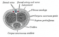 Transverse section of the penis.
 -
The place of the urethral opening in women.
-
 The place of the urethral opening in men.


.jpg)
See also
- Perineal urethra
- Vulvovaginal health
- Urethral sponge
- G-spot
- Sexual stimulation: Urethral sounding and Urethral intercourse
- Urethrotomy
- Urethral Stricture
- Cystoscopy
- Internal urethral orifice
References
- ^ Atlas of Human Anatomy 5th Edition, Netter.
- ^ Zacharin RF (1963). "The suspensory mechanism of the female urethra". J. Anat. 97 (Pt 3): 423–7. PMC 1244203. PMID 14047361.
{{cite journal}}: Unknown parameter|month=ignored (help) - ^ Kohler TS, Yadven M, Manvar A, Liu N, Monga M (2008). "The length of the male urethra". International Braz J Urol. 34 (4): 451–4, discussion 455–6. PMID 18778496.
{{cite journal}}: CS1 maint: multiple names: authors list (link) - ^ Stravodimos, Konstantinos G; Koritsiadis, Georgios; Koutalellis, Georgios (2009). "Electrical wire as a foreign body in a male urethra: a case report". Journal of Medical Case Reports. 3: 49. doi:10.1186/1752-1947-3-49. PMC 2649937. PMID 19192284.
{{cite journal}}: CS1 maint: unflagged free DOI (link)
External links
- Histology at KUMC epithel-epith07 "Male Urethra"