
Pancreas: Difference between revisions
m Reverted edits by 209.174.128.2 to last revision by 193.227.170.13 (HG) |
|||
| Line 78: | Line 78: | ||
==History== |
==History== |
||
The pancreas was first identified by [[Herophilus]] (335-280 BC), a [[Greeks|Greek]] [[anatomist]] and [[surgery|surgeon]]. Only a few hundred years later, [[Ruphos]], another Greek anatomist, gave the pancreas its name. The term "pancreas" is derived from the [[Greek language|Greek]] ''pan'', "all", and ''kreas'', "flesh", probably referring to the organ's homogeneous appearance.<ref>{{cite web |last=Harper |first=Douglas |title=Pancreas |work=Online Etymology Dictionary |url=http://www.etymonline.com/index.php?term=pancreas |accessdate=2007-04-04}}</ref> |
The pancreas was first identified by [[Herophilus]] (335-280 BC), a [[Greeks|Greek]] [[anatomist]] and [[surgery|surgeon]]. Only a few hundred years later, [[Ruphos]], another Greek anatomist, gave the pancreas its name. The term "pancreas" is derived from the [[Greek language|Greek]] ''pan'', "all", and ''kreas'', "flesh", probably referring to the organ's homogeneous appearance.<ref>{{cite web |last=Harper |first=Douglas |title=Pancreas |work=Online Etymology Dictionary |url=http://www.etymonline.com/index.php?term=pancreas |accessdate=2007-04-04}}</ref> your a homo |
||
==Embryological development== |
==Embryological development== |
||
Revision as of 14:01, 28 April 2009


The pancreas is a gland organ in the digestive and endocrine system of vertebrates. It is both an endocrine gland producing several important hormones, including insulin, glucagon, and somatostatin, as well as an exocrine gland, secreting pancreatic juice containing digestive enzymes that pass to the small intestine. These enzymes help in the further breakdown of the carbohydrates, protein, and fat in the chyme.
Histology
Under a microscope, stained sections of the pancreas reveal two different types of parenchymal tissue.[2] Lightly staining clusters of cells are called islets of Langerhans, which produce hormones that underlie the endocrine functions of the pancreas. Darker staining cells form acini connected to ducts. Acinar cells belong to the exocrine pancreas and secrete digestive enzymes into the gut via a system of ducts.
| Structure | Appearance | Function |
| Islets of Langerhans | Lightly staining, large, spherical clusters | Hormone production and secretion (endocrine pancreas) |
| Pancreatic acini | Darker staining, small, berry-like clusters | Digestive enzyme production and secretion (exocrine pancreas) |
Function
The pancreas is a dual-function gland, having features of both endocrine and exocrine glands.
Endocrine
The part of the pancreas with endocrine function is made up of approximately million[3] cell clusters called islets of Langerhans. There are four main cell types in the islets. They are relatively difficult to distinguish using standard staining techniques, but they can be classified by their secretion: α cells secrete glucagon, β cells secrete insulin, δ cells secrete somatostatin, and PP cells secrete pancreatic polypeptide.[4]
The islets are a compact collection of endocrine cells arranged in clusters and cords and are crisscrossed by a dense network of capillaries. The capillaries of the islets are lined by layers of endocrine cells in direct contact with vessels, and most endocrine cells are in direct contact with blood vessels, by either cytoplasmic processes or by direct apposition. According to the volume The Body, by Alan E. Nourse,[5] the islets are "busily manufacturing their hormone and generally disregarding the pancreatic cells all around them, as though they were located in some completely different part of the body."
Exocrine
In contrast to the endocrine pancreas, which secretes hormones into the blood, the exocrine pancreas produces digestive enzymes and an alkaline fluid, and secretes them into the small intestine through a system of exocrine ducts in response to the small intestine hormones secretin and cholecystokinin. Digestive enzymes include trypsin, chymotrypsin, pancreatic lipase, and pancreatic amylase, and are produced and secreted by acinar cells of the exocrine pancreas. Specific cells that line the pancreatic ducts, called centroacinar cells, secrete a bicarbonate- and salt-rich solution into the small intestine.[6]
Regulation
The pancreas receives regulatory innervation via hormones in the blood and through the autonomic nervous system. These two inputs regulate the secretory activity of the pancreas.
| Sympathetic (adrenergic) | Parasympathetic (muscarinic) |
| α2: decreases secretion from beta cells, increases secretion from alpha cells | M3[7] increases stimulation from alpha cells and beta cell |
Diseases of the pancreas
Because the pancreas is a storage depot for digestive enzymes, injury to the pancreas is potentially very dangerous. A puncture of the pancreas generally requires prompt and experienced medical intervention.
An incision into the pancreas is known as a pancreatotomy.
History
The pancreas was first identified by Herophilus (335-280 BC), a Greek anatomist and surgeon. Only a few hundred years later, Ruphos, another Greek anatomist, gave the pancreas its name. The term "pancreas" is derived from the Greek pan, "all", and kreas, "flesh", probably referring to the organ's homogeneous appearance.[8] your a homo
Embryological development

The pancreas forms from the embryonic foregut and is therefore of endodermal origin. Pancreatic development begins the formation of a ventral and dorsal anlage (or buds). Each structure communicates with the foregut through a duct. The ventral pancreatic bud becomes the head and unciate process, and comes from the hepatic diverticulum.
Differential rotation and fusion of the ventral and dorsal pancreatic buds results in the formation of the definitive pancreas.[9] As the duodenum rotates to the right, it carries with it the ventral pancreatic bud and common bile duct. Upon reaching its final destination, the ventral pancreatic bud fuses with the much larger dorsal pancreatic bud. At this point of fusion, the main ducts of the ventral and dorsal pancreatic buds fuse, forming the duct of Wirsung, the main pancreatic duct.
Differentiation of cells of the pancreas proceeds through two different pathways, corresponding to the dual endocrine and exocrine functions of the pancreas. In progenitor cells of the exocrine pancreas, important molecules that induce differentiation include follistatin, fibroblast growth factors, and activation of the Notch receptor system.[9] Development of the exocrine acini progresses through three successive stages. These include the predifferentiated, protodifferentiated, and differentiated stages, which correspond to undetectable, low, and high levels of digestive enzyme activity, respectively.
Progenitor cells of the endocrine pancreas arise from cells of the protodifferentiated stage of the exocrine pancreas.[9] Under the influence of neurogenin-3 and Isl-1, but in the absence of Notch receptor signaling, these cells differentiate to form two lines of committed endocrine precursor cells. The first line, under the direction of Pax-0, forms α- and γ- cells, which produce the peptides glucagon and pancreatic polypeptide, respectively. The second line, influenced by Pax-6, produces β- and δ-cells, which secrete insulin and somatostatin, respectively.
Insulin and glucagon can be detected in the fetal circulation by the fourth or fifth month of fetal development.[9]
As food
The pancreas of lamb, beef, and pork is prepared and eaten. Along with the thymus, pancreas is considered a sweetbread.
Additional images
-
 Accessory digestive system.
 -
 Digestive organs.
 -
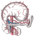 The celiac artery and its branches; the stomach has been raised and the peritoneum removed.
 -
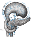 Lymphatics of stomach, etc. The stomach has been turned upward.
 -
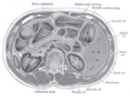 Transverse section through the middle of the first lumbar vertebra, showing the relations of the pancreas.
 -
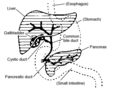
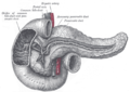
 The duodenum and pancreas.
 -
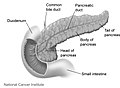
 The pancreatic duct.
 -
 Pancreas of a human embryo of five weeks.
 -
 Pancreas of a human embryo at end of sixth week.
 -
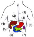
 Front of abdomen, showing surface markings for duodenum, pancreas, and kidneys.
 -


References
- ^ Template:GeorgiaPhysiology
- ^ Histology image: 10404loa – Histology Learning System at Boston University
- ^ Hellman B, Gylfe E, Grapengiesser E, Dansk H, Salehi A (2007). "[Insulin oscillations--clinically important rhythm. Anti-diabetics should increase the pulsative component of the insulin release]". Lakartidningen (in Swedish). 104 (32–33): 2236–9. PMID 17822201.
{{cite journal}}: CS1 maint: multiple names: authors list (link) - ^ BRS physiology 4th edition ,page 255-256, Linda S. Constanzo, Lippincott publishing
- ^ The Body, by Alan E. Nourse, in the Time-Life Science Library Series (op. cit., p. 171.)
- ^ Maton, Anthea (1993). Human Biology and Health. Englewood Cliffs, New Jersey, USA: Prentice Hall. ISBN 0-13-981176-1. OCLC 32308337.
{{cite book}}: Unknown parameter|coauthors=ignored (|author=suggested) (help) - ^ Verspohl EJ, Tacke R, Mutschler E, Lambrecht G (1990). "Muscarinic receptor subtypes in rat pancreatic islets: binding and functional studies". Eur. J. Pharmacol. 178 (3): 303–11. doi:10.1016/0014-2999(90)90109-J. PMID 2187704.
{{cite journal}}: CS1 maint: multiple names: authors list (link) - ^ Harper, Douglas. "Pancreas". Online Etymology Dictionary. Retrieved 2007-04-04.
- ^ a b c d Carlson, Bruce M. (2004). Human embryology and developmental biology. St. Louis: Mosby. pp. 372–4. ISBN 0-323-01487-9.